Submandibular lymph nodes: Difference between revisions
No edit summary |
→Additional images: lowercase |
||
| (3 intermediate revisions by 2 users not shown) | |||
| Line 1: | Line 1: | ||
{{Short description|Lymph nodes near the jaw}} |
|||
{{Infobox lymph |
{{Infobox lymph |
||
| Name = Submandibular lymph nodes |
| Name = Submandibular lymph nodes |
||
| Latin = |
| Latin = nodi lymphoidei submandibulares |
||
| Image = Illu quiz hn 03.jpg |
| Image = Illu quiz hn 03.jpg |
||
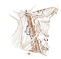
| Caption = 1: [[Submental lymph nodes]]<br />2: Submandibular lymph nodes<br />3: [[Supraclavicular lymph nodes]]<br />4: [[Retropharyngeal lymph nodes]]<br />5: [[Buccinator lymph node]]<br />6: [[Superficial cervical lymph nodes]]<br />7: [[Jugular lymph nodes]]<br />8: [[Parotid lymph nodes]]<br />9: [[Retroauricular lymph nodes]] |
| Caption = 1: [[Submental lymph nodes]]<br />2: Submandibular lymph nodes<br />3: [[Supraclavicular lymph nodes]]<br />4: [[Retropharyngeal lymph nodes]]<br />5: [[Buccinator lymph node]]<br />6: [[Superficial cervical lymph nodes]]<br />7: [[Jugular lymph nodes]]<br />8: [[Parotid lymph nodes]]<br />9: [[Retroauricular lymph nodes]] and [[occipital lymph nodes]] |
||
| Image2 = Gray602.png |
| Image2 = Gray602.png |
||
| Caption2 = Superficial lymph glands and lymphatic vessels of head and neck. (Submaxillary glands labeled at center right.) |
| Caption2 = Superficial lymph glands and lymphatic vessels of head and neck. (Submaxillary glands labeled at center right.) |
||
| Line 32: | Line 33: | ||
== Additional images == |
== Additional images == |
||
<gallery> |
<gallery> |
||
File:illu_lymph_chain02.jpg|Deep |
File:illu_lymph_chain02.jpg|Deep lymph nodes |
||
</gallery> |
</gallery> |
||
Latest revision as of 15:19, 16 May 2024
| Submandibular lymph nodes | |
|---|---|
 1: Submental lymph nodes 2: Submandibular lymph nodes 3: Supraclavicular lymph nodes 4: Retropharyngeal lymph nodes 5: Buccinator lymph node 6: Superficial cervical lymph nodes 7: Jugular lymph nodes 8: Parotid lymph nodes 9: Retroauricular lymph nodes and occipital lymph nodes | |
 Superficial lymph glands and lymphatic vessels of head and neck. (Submaxillary glands labeled at center right.) | |
| Details | |
| System | Lymphatic system |
| Source | Mandibular lymph node |
| Identifiers | |
| Latin | nodi lymphoidei submandibulares |
| Anatomical terminology | |
The submandibular lymph nodes (submaxillary glands in older texts), are some 3-6 lymph nodes situated at the inferior border of the ramus of mandible.[1]
Anatomy
[edit]They are situated just superficial to the submandibular salivary gland, and posterolateral to the anterior belly of either digastric muscle.[1]
One gland, the middle gland of Stahr, which lies on the facial artery as it turns over the mandible, is the most constant of the series; small lymph glands are sometimes found on the deep surface of the submandibular gland.[citation needed]
Afferents
[edit]They drain the upper lip, body of tongue, cheeks, anterior portion of the hard palate, and most teeth with their associated periodontium and gingiva (except for the mandibular incisor teeth and third molar teeth).[1]
The facial and submental lymph nodes may also drain into the submandibular glands.[1]
Efferents
[edit]They drain to the superior[citation needed] deep cervical lymph nodes.[1]
Clinical significance
[edit]The most common causes of enlargement of the submandibular lymph nodes are infections of the head, neck, ears, eyes, nasal sinuses, pharynx, and scalp.[1]
The lymph glands may be affected by metastatic spread of cancers of the oral cavity, anterior portion of the nasal cavity, soft tissues of the mid-face, and submandibular salivary gland.[1]
Additional images
[edit]-
Deep lymph nodes
References
[edit]![]() This article incorporates text in the public domain from page 697 of the 20th edition of Gray's Anatomy (1918)
This article incorporates text in the public domain from page 697 of the 20th edition of Gray's Anatomy (1918)
External links
[edit]- Archived Diagram via umich.edu - rollover to see labels
- https://web.archive.org/web/20080216031919/http://www.med.mun.ca/anatomyts/head/hnl3a.htm
- Diagram at Baylor College of Medicine
- http://www.patient.info/
- http://www.aafp.org/afp/20021201/2103.html
- http://www.emedicine.com/ent/topic306.htm#section~anatomy_of_the_cervical_lymphatics

