Nodular melanoma: Difference between revisions
No edit summary |
cite |
||
| (11 intermediate revisions by 9 users not shown) | |||
| Line 20: | Line 20: | ||
}} |
}} |
||
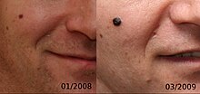
[[Image:NodularMelanomaEvolution.jpg|thumb|alt=Evolution of a 4 mm nodular melanoma.|Evolution of a 4 mm nodular melanoma.]] |
[[Image:NodularMelanomaEvolution.jpg|thumb|alt=Evolution of a 4 mm nodular melanoma.|Evolution of a 4 mm nodular melanoma.]] |
||
'''Nodular melanoma''' ('''NM''') is the most aggressive form of [[melanoma]]. It tends to grow more rapidly in thickness (penetrate the skin) than in diameter. Instead of arising from a pre-existing mole, it may appear in a spot where a lesion did not previously exist. Since NM tends to grow in depth more quickly than it does in width, and can occur in a place that did not have a previous lesion, the prognosis is often worse because it takes longer for a person to be aware of the changes. NM is most often darkly pigmented; however, some NM lesions can be light brown, multicolored or even colorless (non-pigmented). A light-colored or non-pigmented NM lesion may escape detection because the appearance is not alarming, however an ulcerated and/or bleeding lesion is common.<ref name="Andrews">{{cite book |author=James, William D. |author2=Berger, Timothy G.|title=Andrews' Diseases of the Skin: clinical Dermatology |publisher=Saunders Elsevier |
'''Nodular melanoma''' ('''NM''') is the most aggressive form of [[melanoma]].<ref name=WHO2018>{{cite book |last1=DE |first1=Elder |last2=D |first2=Massi |last3=RA |first3=Scolyer |last4=R |first4=Willemze |title=WHO Classification of Skin Tumours |date=2018 |publisher=World Health Organization |location=Lyon (France) |isbn=978-92-832-2440-2 |edition=4th |volume=11 |url=https://publications.iarc.fr/Book-And-Report-Series/Who-Classification-Of-Tumours/WHO-Classification-Of-Skin-Tumours-2018 |language=en |chapter=2. Melanocytic tumours |pages=145-146 }}</ref> It tends to grow more rapidly in thickness (vertically penetrate the skin) than in diameter compared to other melanoma subtypes.<ref name=":0">{{Cite journal|last1=Egger|first1=Michael E.|last2=Dunki-Jacobs|first2=Erik M.|last3=Callender|first3=Glenda G.|last4=Quillo|first4=Amy R.|last5=Scoggins|first5=Charles R.|last6=Martin|first6=Robert C.G.|last7=Stromberg|first7=Arnold J.|last8=McMasters|first8=Kelly M.|date=October 2012|title=Outcomes and prognostic factors in nodular melanomas|journal=Surgery|volume=152|issue=4|pages=652–660|doi=10.1016/j.surg.2012.07.006|pmid=22925134|issn=0039-6060}}</ref> Instead of arising from a pre-existing mole, it may appear in a spot where a lesion did not previously exist. Since NM tends to grow in depth more quickly than it does in width, and can occur in a place that did not have a previous lesion, the prognosis is often worse because it takes longer for a person to be aware of the changes. NM is most often darkly pigmented; however, some NM lesions can be light brown, multicolored or even colorless (non-pigmented). A light-colored or non-pigmented NM lesion may escape detection because the appearance is not alarming, however an ulcerated and/or bleeding lesion is common.<ref name="Andrews">{{cite book |author=James, William D. |author2=Berger, Timothy G.|title=Andrews' Diseases of the Skin: clinical Dermatology |publisher=Saunders Elsevier |year=2006 |isbn=978-0-7216-2921-6 |display-authors=etal}}</ref>{{rp|696}} [[Polypoid melanoma]] is a virulent variant of nodular melanoma.<ref name="Andrews"/>{{rp|696}} |
||
The microscopic hallmarks are: |
The microscopic hallmarks are: |
||
| Line 27: | Line 27: | ||
* Dermal nodule of melanocytes with a 'pushing' growth pattern |
* Dermal nodule of melanocytes with a 'pushing' growth pattern |
||
* No "radial growth phase" |
* No "radial growth phase" |
||
__NOTOC__ |
|||
==Treatment== |
==Treatment== |
||
Therapies for metastatic melanoma include the biologic immunotherapy agents [[ipilimumab]], [[pembrolizumab]], and [[nivolumab]]; [[BRAF inhibitor]]s, such as [[vemurafenib]] and [[dabrafenib]]; and a [[MEK inhibitor]] [[trametinib]].<ref>{{cite journal | vauthors = Maverakis E, Cornelius LA, Bowen GM, Phan T, Patel FB, Fitzmaurice S, He Y, Burrall B, Duong C, Kloxin AM, Sultani H, Wilken R, Martinez SR, Patel F | title = Metastatic melanoma - a review of current and future treatment options | journal = Acta Derm Venereol | volume = 95 | issue = 5 | pages = 516–524 | year = 2015 | pmid = 25520039 | doi = 10.2340/00015555-2035| url = http://digitalcommons.wustl.edu/cgi/viewcontent.cgi?article=4849&context=open_access_pubs | doi-access = free }}</ref> |
Therapies for metastatic melanoma include the biologic immunotherapy agents [[ipilimumab]], [[pembrolizumab]], and [[nivolumab]]; [[BRAF inhibitor]]s, such as [[vemurafenib]] and [[dabrafenib]]; and a [[MEK inhibitor]] [[trametinib]].<ref>{{cite journal | vauthors = Maverakis E, Cornelius LA, Bowen GM, Phan T, Patel FB, Fitzmaurice S, He Y, Burrall B, Duong C, Kloxin AM, Sultani H, Wilken R, Martinez SR, Patel F | title = Metastatic melanoma - a review of current and future treatment options | journal = Acta Derm Venereol | volume = 95 | issue = 5 | pages = 516–524 | year = 2015 | pmid = 25520039 | doi = 10.2340/00015555-2035| url = http://digitalcommons.wustl.edu/cgi/viewcontent.cgi?article=4849&context=open_access_pubs | doi-access = free }}</ref> |
||
== Prognosis == |
|||
Important prognosis factors for nodular melanoma include: |
|||
* Thickness |
|||
* Ulceration |
|||
* [[Sentinel lymph node|Sentinel lymph node (SLN)]] status<ref name=":0" /> |
|||
== See also == |
== See also == |
||
Latest revision as of 15:36, 21 May 2024
| Nodular melanoma | |
|---|---|
 | |
| Specialty | Oncology, dermatology |

Nodular melanoma (NM) is the most aggressive form of melanoma.[1] It tends to grow more rapidly in thickness (vertically penetrate the skin) than in diameter compared to other melanoma subtypes.[2] Instead of arising from a pre-existing mole, it may appear in a spot where a lesion did not previously exist. Since NM tends to grow in depth more quickly than it does in width, and can occur in a place that did not have a previous lesion, the prognosis is often worse because it takes longer for a person to be aware of the changes. NM is most often darkly pigmented; however, some NM lesions can be light brown, multicolored or even colorless (non-pigmented). A light-colored or non-pigmented NM lesion may escape detection because the appearance is not alarming, however an ulcerated and/or bleeding lesion is common.[3]: 696 Polypoid melanoma is a virulent variant of nodular melanoma.[3]: 696
The microscopic hallmarks are:
- Dome-shaped at low power
- Epidermis thin or normal
- Dermal nodule of melanocytes with a 'pushing' growth pattern
- No "radial growth phase"
Treatment
[edit]Therapies for metastatic melanoma include the biologic immunotherapy agents ipilimumab, pembrolizumab, and nivolumab; BRAF inhibitors, such as vemurafenib and dabrafenib; and a MEK inhibitor trametinib.[4]
Prognosis
[edit]Important prognosis factors for nodular melanoma include:
- Thickness
- Ulceration
- Sentinel lymph node (SLN) status[2]
See also
[edit]References
[edit]- ^ DE, Elder; D, Massi; RA, Scolyer; R, Willemze (2018). "2. Melanocytic tumours". WHO Classification of Skin Tumours. Vol. 11 (4th ed.). Lyon (France): World Health Organization. pp. 145–146. ISBN 978-92-832-2440-2.
- ^ a b Egger, Michael E.; Dunki-Jacobs, Erik M.; Callender, Glenda G.; Quillo, Amy R.; Scoggins, Charles R.; Martin, Robert C.G.; Stromberg, Arnold J.; McMasters, Kelly M. (October 2012). "Outcomes and prognostic factors in nodular melanomas". Surgery. 152 (4): 652–660. doi:10.1016/j.surg.2012.07.006. ISSN 0039-6060. PMID 22925134.
- ^ a b James, William D.; Berger, Timothy G.; et al. (2006). Andrews' Diseases of the Skin: clinical Dermatology. Saunders Elsevier. ISBN 978-0-7216-2921-6.
- ^ Maverakis E, Cornelius LA, Bowen GM, Phan T, Patel FB, Fitzmaurice S, He Y, Burrall B, Duong C, Kloxin AM, Sultani H, Wilken R, Martinez SR, Patel F (2015). "Metastatic melanoma - a review of current and future treatment options". Acta Derm Venereol. 95 (5): 516–524. doi:10.2340/00015555-2035. PMID 25520039.
