Stratified cuboidal epithelium: Difference between revisions
Appearance
Content deleted Content added
No edit summary |
Adding local short description: "Tissue made of cube-shaped cells", overriding Wikidata description "tissue type" |
||
| (20 intermediate revisions by 19 users not shown) | |||
| Line 1: | Line 1: | ||
{{Short description|Tissue made of cube-shaped cells}} |
|||
{{Infobox |
{{Infobox cell |
||
| Name = Stratified cuboidal epithelium |
| Name = Stratified cuboidal epithelium |
||
| Latin = Epithelium stratificatum cuboideum |
| Latin = Epithelium stratificatum cuboideum |
||
| Greek = |
| Greek = |
||
| GraySubject = |
|||
| GrayPage = |
|||
| Image = WVSOM Parotid Gland1.JPG |
| Image = WVSOM Parotid Gland1.JPG |
||
| Caption = Stratified cuboidal epithelium in the ducts of the [[parotid gland]], visible as the borders of the two circular structures in the upper left. |
| Caption = Stratified cuboidal epithelium in the ducts of the [[parotid gland]], visible as the borders of the two circular structures in the upper left. |
||
| Line 10: | Line 9: | ||
| Image2 = |
| Image2 = |
||
| Caption2 = |
| Caption2 = |
||
| ImageMap = |
|||
| MapCaption = |
|||
| Precursor = |
| Precursor = |
||
| |
| Shape = Many layers of cuboid cells |
||
| Artery = |
|||
| Vein = |
|||
| Nerve = |
|||
| Lymph = |
|||
| MeshName = |
|||
| MeshNumber = |
|||
| Code = {{TerminologiaHistologica|2|00|02.0.02031}} |
|||
| Dorlands = |
|||
| DorlandsID = |
|||
}} |
}} |
||
{{Epithelia series}} |
{{Epithelia series}} |
||
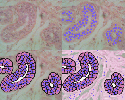
[[File:Stratified cuboidal epithelium animated.gif|alt=Stratified cuboidal epithelium|thumb|Stratified cuboidal epithelium, highlighting the nucleuses, the rest of the epithelial cells, and underlying connective tissue.]] |
|||
'''Stratified cuboidal epithelium''' is a type of [[epithelial tissue]] composed of multiple layers of cube-shaped cells. Only the most superficial layer is made up of cuboidal cells, and the other layers can be cells of other types. Topmost layer of [[skin]] [[epidermis]] in [[Frog|frogs]], [[fish]] is made up of living cuboidal cells. |
|||
==Structure== |
|||
Only the most superficial layer is made up of cuboidal cells, and the other layers can be cells of other types. |
|||
| ⚫ | This type of tissue can be observed in [[sweat gland]]s, [[mammary gland]]s, [[circumanal gland]]s, and [[salivary gland]]s.<ref name=KMU1>{{cite web|url=http://hist.vexp.idv.tw/class/histo01.htm|title=Epithelium|publisher=KMU(高雄醫學大學)|language=Chinese}}</ref><ref>{{cite web|url=http://hist.class.kmu.edu.tw/Basic/Epithelial/Stratified/Cuboidal/index.html|title=Stratified cuboidal epithelium|publisher=KMU(高雄醫學大學)|language=Chinese}}</ref> They protect areas such as the ducts of [[sweat gland]]s,<ref name=eroschenko08>{{cite book| publisher = Lippincott Williams & Wilkins| isbn = 9780781770576| pages = [https://archive.org/details/difioresatlashis00eros/page/n231 212]–234| last = Eroschenko| first = Victor P.| title = DiFiore's Atlas of Histology with Functional Correlations| url = https://archive.org/details/difioresatlashis00eros| url-access = limited| chapter = Integumentary System| year = 2008}}</ref> [[mammary gland]]s, and [[salivary gland]]s. They are also observed in the linings of [[urethra]]. |
||
==Distribution and Function== |
|||
| ⚫ | This type of tissue can be observed in [[sweat gland]]s, [[mammary gland]]s, [[ |
||
==References== |
==References== |
||
| Line 36: | Line 23: | ||
==External links== |
==External links== |
||
* [http://www3.umdnj.edu/histsweb/lab2/lab2stratifiedcuboidal.html Overview at umdnj.edu] |
* [https://web.archive.org/web/20060919140049/http://www3.umdnj.edu/histsweb/lab2/lab2stratifiedcuboidal.html Overview at umdnj.edu] |
||
* {{KansasHistology|epithel|epith17}} "Sweat Duct" (Stratified cuboidal) |
* {{KansasHistology|epithel|epith17}} "Sweat Duct" (Stratified cuboidal) |
||
* {{OklahomaHistology|43_03}} - "Skin" |
* {{OklahomaHistology|43_03}} - "Skin" |
||
{{Epithelium and epithelial tissue}} |
{{Epithelium and epithelial tissue}} |
||
{{Authority control}} |
|||
[[Category:Epithelium]] |
[[Category:Epithelium]] |
||
Latest revision as of 01:26, 19 October 2024
| Stratified cuboidal epithelium | |
|---|---|
 Stratified cuboidal epithelium in the ducts of the parotid gland, visible as the borders of the two circular structures in the upper left. | |
| Details | |
| Shape | Many layers of cuboid cells |
| Identifiers | |
| Latin | Epithelium stratificatum cuboideum |
| TH | H2.00.02.0.02031 |
| FMA | 63912 |
| Anatomical terms of microanatomy | |
| This article is part of a series on |
| Epithelia |
|---|
| Squamous epithelial cell |
| Columnar epithelial cell |
| Cuboidal epithelial cell |
| Specialised epithelia |
|
| Other |

Stratified cuboidal epithelium is a type of epithelial tissue composed of multiple layers of cube-shaped cells. Only the most superficial layer is made up of cuboidal cells, and the other layers can be cells of other types. Topmost layer of skin epidermis in frogs, fish is made up of living cuboidal cells.
Structure
[edit]This type of tissue can be observed in sweat glands, mammary glands, circumanal glands, and salivary glands.[1][2] They protect areas such as the ducts of sweat glands,[3] mammary glands, and salivary glands. They are also observed in the linings of urethra.
References
[edit]- ^ "Epithelium" (in Chinese). KMU(高雄醫學大學).
- ^ "Stratified cuboidal epithelium" (in Chinese). KMU(高雄醫學大學).
- ^ Eroschenko, Victor P. (2008). "Integumentary System". DiFiore's Atlas of Histology with Functional Correlations. Lippincott Williams & Wilkins. pp. 212–234. ISBN 9780781770576.
External links
[edit]- Overview at umdnj.edu
- Histology at KUMC epithel-epith17 "Sweat Duct" (Stratified cuboidal)
- Histology image: 43_03 at the University of Oklahoma Health Sciences Center - "Skin"
