Pedipalp: Difference between revisions
text layout; minor citation touchups (links, etc); caption edits; |
Reverting edit(s) by 120.29.78.137 (talk) to rev. 1262810552 by Mrfoogles: Not providing a reliable source (RW 16.1) |
||
| (14 intermediate revisions by 13 users not shown) | |||
| Line 1: | Line 1: | ||
{{Short description|Appendage of chelicerate}} |
{{Short description|Appendage of chelicerate}} |
||
{{about|chelicerate pedipalps |
{{about|chelicerate pedipalps}} |
||
| ⚫ | |||
{{multiple image |
|||
|total_width=400 |
|||
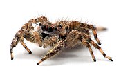
| ⚫ | '''Pedipalps''' (commonly shortened to '''palps''' or '''palpi''') are the secondary pair of forward [[appendage]]s among [[Chelicerata|chelicerates]] – a group of [[arthropod]]s including [[spider]]s, [[scorpion]]s, [[horseshoe crab]]s, and [[sea spider]]s. The pedipalps are lateral to the [[chelicerae]] ("jaws") and anterior to the first pair of walking legs. |
||
|width1=750 |height1=1000 |
|||
|image1=Oxyopes salticus Kaldari 05 crop.jpg |
|||
|caption1=Male [[Oxyopes salticus|striped lynx spider]] showing enlarged pedipalps |
|||
|width2=750 |height2=1120 |
|||
|image2=Pedipalp in green.png |
|||
| ⚫ | |||
}} |
|||
| ⚫ | '''Pedipalps''' (commonly shortened to '''palps''' or '''palpi''') are the |
||
==Overview== |
==Overview== |
||
[[File:Zoropsis spinimana-pjt2.jpg|thumb|right|Male [[Zoropsis spinimana]] showing enlarged pedipalps]] |
|||
Pedipalps are composed of six segments or articles: the coxa, the [[Arthropod leg# |
Pedipalps are composed of six segments or articles. From the proximal end (where they are attached to the spider) to the distal, they are: the coxa, the [[Arthropod leg#Trochanter|trochanter]], the [[Arthropod leg#Femur|femur]], the short [[Glossary_of_spider_terms#patella|patella]], the [[Glossary_of_spider_terms#tibia|tibia]], and the [[Arthropod_leg#Tarsus|tarsus]]. In spiders, the coxae frequently have extensions called [[Glossary_of_spider_terms#maxilla |maxillae]] or gnathobases, which function as mouth parts with or without some contribution from the coxae of the anterior [[arthropod leg|legs]]. The limbs themselves may be simple tactile organs outwardly resembling the legs, as in [[spider]]s, or [[Chela (organ)|chelate]] weapons (pincers) of great size, as in [[scorpion]]s. The pedipalps of [[Solifugae]] are covered in [[seta]]e, but have not been studied in detail.<ref>{{cite journal |last1=Cushing |first1=Paula |last2=Casto |first2=Patrick |date=2012 |title=Preliminary survey of the setal and sensory structures on the pedipalps of camel spiders (Arachnida: Solifugae) |journal=[[The Journal of Arachnology]] |volume=40 |issue=1 |pages=123–127 |doi=10.1636/B11-71.1 |s2cid=86837385 |url=https://bioone.org/journals/The-Journal-of-Arachnology/volume-40/issue-1/B11-71.1/Preliminary-survey-of-the-setal-and-sensory-structures-on-the/10.1636/B11-71.1.full |access-date=5 September 2020}}</ref> |
||
Comparative studies of pedipalpal morphology may suggest that leg-like pedipalps are [[primitive (biology)|primitive]] in arachnids. At present, the only reasonable alternative to this view{{according to whom?|date=February 2016}} is to assume that [[ |
Comparative studies of pedipalpal morphology may suggest that leg-like pedipalps are [[primitive (biology)|primitive]] in arachnids. At present, the only reasonable alternative to this view{{according to whom?|date=February 2016}} is to assume that [[xiphosura]]ns reflect the morphology of the primitive arachnid pedipalp and to conclude that this appendage is primitively chelate. Pedipalps are traditionally thought to be [[Homology (biology)|homologous]] with [[Mandible (arthropod)|mandibles]] in [[crustaceans]] and [[insect]]s, although more recent studies (e.g. using [[Hox genes]]) suggest they are probably homologous with the crustacean second [[antennae (biology)|antennae]]. |
||
==Chelate pedipalps== |
==Chelate pedipalps== |
||
| Line 25: | Line 19: | ||
[[File:Unicorn catleyi copulatory bulb.png|thumb|Palpal or copulatory bulb of [[Unicorn (genus)|''Unicorn catleyi'']]]] |
[[File:Unicorn catleyi copulatory bulb.png|thumb|Palpal or copulatory bulb of [[Unicorn (genus)|''Unicorn catleyi'']]]] |
||
Pedipalps of [[spiders]] have the same segmentation as the legs, but the [[Spider anatomy|tarsus]] is undivided, and the pretarsus has no lateral claws. Pedipalps contain sensitive chemical detectors and function as taste and smell organs, supplementing those on the legs.<ref>{{ |
Pedipalps of [[spiders]] have the same segmentation as the legs, but the [[Spider anatomy|tarsus]] is undivided, and the pretarsus has no lateral claws. Pedipalps contain sensitive chemical detectors and function as taste and smell organs, supplementing those on the legs.<ref>{{cite web|place=Washington, DC|publisher=[[Smithsonian Institution]]|series=Under the spell of spiders|title=Spider specifics|type=lesson plan|url=http://www.smithsonianeducation.org/educators/lesson_plans/under_spell_spiders/spiderspecifics.html|website=Smithsonian Education}}</ref> In [[sexually mature|sexually-mature]] male spiders, the final segment of the pedipalp, the tarsus, develops a complicated structure (sometimes called the [[palpal bulb]] or palpal organ) that is used to transfer sperm to the female seminal receptacles during mating. The details of this structure vary considerably between different groups of spiders and are useful for identifying species.<ref name="comstock">{{cite book |first=John Henry |last=Comstock |year=1920 |orig-date=1912 |title=The Spider Book |publisher=Doubleday, Page & Company |pages=106–121}}</ref><ref name="foelix">{{cite book |first=Rainer F. |last=Foelix |year=1996 |title=Biology of Spiders |edition=2 |publisher=Oxford University Press |isbn=978-0-19-509594-4 |pages=[https://archive.org/details/biologyofspiders00foel_0/page/182 182–185] |url=https://archive.org/details/biologyofspiders00foel_0 |url-access=registration |via=Internet Archive (archive.org) }}</ref> The pedipalps are also used by male spiders in courtship displays, contributing to vibratory patterns in web-shaking, acoustic signals, or visual displays.<ref>{{cite journal |last1=Pechmann |first1=Matthias |last2=Khadjeh |first2=Sara |last3=Sprenger |first3=Fredrik |last4=Prpic |first4=Nikola-Michael |date=November 2010 |title=Patterning mechanisms and morphological diversity of spider appendages and their importance for spider evolution |journal=Arthropod Structure & Development |volume=39 |issue=6 |pages=453–467 |doi=10.1016/j.asd.2010.07.007 |pmid=20696272 |url=https://www.sciencedirect.com/science/article/pii/S1467803910000551 |access-date=20 August 2020}}</ref> |
||
The '''[[palpal bulb|cymbium]]''' is a spoon-shaped structure located at the end of the spider pedipalp that supports the palpal organ.<ref name=comstock/> The cymbium may also be used as a [[stridulatory organ]] in spider courtship.<ref>{{cite |
The '''[[palpal bulb|cymbium]]''' is a spoon-shaped structure located at the end of the spider pedipalp that supports the palpal organ.<ref name=comstock/> The cymbium may also be used as a [[stridulatory organ]] in spider courtship.<ref>{{cite journal |author1=Elias, Damian O. |author2=Lee, Norman |author3=Hebets, Eileen A. |author4=Mason, Andrew C. |date=15 March 2006 |title=Seismic signal production in a wolf spider: Parallel versus serial multi-component signals |journal=[[Journal of Experimental Biology]] |volume=209 |issue=6 |pages=1074–1084 |doi=10.1242/jeb.02104 |pmid=16513934 |doi-access=free}} See [http://jeb.biologists.org/cgi/content/full/209/6/1074/FIG6 Fig 6 – SEM of tibio-cymbial joint on the male palp of ''S. stridulans'']</ref> |
||
The '''embolus''' is a narrow whip-like or leaf-like extension of the palpal bulb. |
The '''embolus''' is a narrow whip-like or leaf-like extension of the palpal bulb. |
||
Latest revision as of 14:41, 23 December 2024

Pedipalps (commonly shortened to palps or palpi) are the secondary pair of forward appendages among chelicerates – a group of arthropods including spiders, scorpions, horseshoe crabs, and sea spiders. The pedipalps are lateral to the chelicerae ("jaws") and anterior to the first pair of walking legs.
Overview
[edit]
Pedipalps are composed of six segments or articles. From the proximal end (where they are attached to the spider) to the distal, they are: the coxa, the trochanter, the femur, the short patella, the tibia, and the tarsus. In spiders, the coxae frequently have extensions called maxillae or gnathobases, which function as mouth parts with or without some contribution from the coxae of the anterior legs. The limbs themselves may be simple tactile organs outwardly resembling the legs, as in spiders, or chelate weapons (pincers) of great size, as in scorpions. The pedipalps of Solifugae are covered in setae, but have not been studied in detail.[1]
Comparative studies of pedipalpal morphology may suggest that leg-like pedipalps are primitive in arachnids. At present, the only reasonable alternative to this view[according to whom?] is to assume that xiphosurans reflect the morphology of the primitive arachnid pedipalp and to conclude that this appendage is primitively chelate. Pedipalps are traditionally thought to be homologous with mandibles in crustaceans and insects, although more recent studies (e.g. using Hox genes) suggest they are probably homologous with the crustacean second antennae.
Chelate pedipalps
[edit]Chelate or sub-chelate (pincer-like) pedipalps are found in several arachnid groups (Ricinulei, Uropygi, scorpions and pseudoscorpions) but the chelae in most of these groups may not be homologous with those found in Xiphosura. The pedipalps are distinctly raptorial (i.e., modified for seizing prey) in the Amblypygi, Uropygi, Schizomida, and some Opiliones belonging to the laniatorid group.[citation needed]
Spider pedipalps
[edit]
Pedipalps of spiders have the same segmentation as the legs, but the tarsus is undivided, and the pretarsus has no lateral claws. Pedipalps contain sensitive chemical detectors and function as taste and smell organs, supplementing those on the legs.[2] In sexually-mature male spiders, the final segment of the pedipalp, the tarsus, develops a complicated structure (sometimes called the palpal bulb or palpal organ) that is used to transfer sperm to the female seminal receptacles during mating. The details of this structure vary considerably between different groups of spiders and are useful for identifying species.[3][4] The pedipalps are also used by male spiders in courtship displays, contributing to vibratory patterns in web-shaking, acoustic signals, or visual displays.[5]
The cymbium is a spoon-shaped structure located at the end of the spider pedipalp that supports the palpal organ.[3] The cymbium may also be used as a stridulatory organ in spider courtship.[6]
The embolus is a narrow whip-like or leaf-like extension of the palpal bulb.
References
[edit]- ^ Cushing, Paula; Casto, Patrick (2012). "Preliminary survey of the setal and sensory structures on the pedipalps of camel spiders (Arachnida: Solifugae)". The Journal of Arachnology. 40 (1): 123–127. doi:10.1636/B11-71.1. S2CID 86837385. Retrieved 5 September 2020.
- ^ "Spider specifics". Smithsonian Education (lesson plan). Under the spell of spiders. Washington, DC: Smithsonian Institution.
- ^ a b Comstock, John Henry (1920) [1912]. The Spider Book. Doubleday, Page & Company. pp. 106–121.
- ^ Foelix, Rainer F. (1996). Biology of Spiders (2 ed.). Oxford University Press. pp. 182–185. ISBN 978-0-19-509594-4 – via Internet Archive (archive.org).
- ^ Pechmann, Matthias; Khadjeh, Sara; Sprenger, Fredrik; Prpic, Nikola-Michael (November 2010). "Patterning mechanisms and morphological diversity of spider appendages and their importance for spider evolution". Arthropod Structure & Development. 39 (6): 453–467. doi:10.1016/j.asd.2010.07.007. PMID 20696272. Retrieved 20 August 2020.
- ^ Elias, Damian O.; Lee, Norman; Hebets, Eileen A.; Mason, Andrew C. (15 March 2006). "Seismic signal production in a wolf spider: Parallel versus serial multi-component signals". Journal of Experimental Biology. 209 (6): 1074–1084. doi:10.1242/jeb.02104. PMID 16513934. See Fig 6 – SEM of tibio-cymbial joint on the male palp of S. stridulans
Further reading
[edit]- Savory, T. (1977). Arachnida (2nd ed.). Academic Press. ISBN 0-12-619660-5.
- Snodgrass, R. E. (1971). A Textbook Arthropod Anatomy. Hafner. OCLC 299721312.
- Torre-Bueno, J.R., ed. (1989). The Torre-Bueno Glossary of Entomology. Nichols, Stephen W. (compiled by); Tulloch, George S. (prepared Supplement A). The New York Entomological Society in cooperation with the American Museum of Natural History.
External links
[edit] Media related to Pedipalps at Wikimedia Commons
- Bokma, John (photographer) (2005-09-07). Brachypelma vagans (Mexican red-rump) (photographs). Veracruz, MX. — Several close-up photos of a tarantula creating a sperm web
- Phrynus longipes#Pedipalps

