Lateral umbilical fold: Difference between revisions
No edit summary |
Citation bot (talk | contribs) Alter: publisher. Removed parameters. | Use this bot. Report bugs. | Suggested by Abductive | #UCB_webform 345/3844 |
||
| (14 intermediate revisions by 13 users not shown) | |||
| Line 1: | Line 1: | ||
{{Infobox |
{{Infobox anatomy |
||
| Name = Lateral umbilical ligament |
|||
| Latin = plica umbilicalis lateralis; plica epigastrica |
|||
| Image = Gray1036.png |
|||
GraySubject = 246 | |
|||
| ⚫ | |||
GrayPage = 1152 | |
|||
| Image2 = Gray1037.png |
|||
| ⚫ | |||
| ⚫ | |||
| System = |
|||
| ⚫ | |||
| ⚫ | |||
}}The '''lateral umbilical fold''' is an elevation (on either side of the body) of the peritoneum lining the inner/posterior surface of the lower [[anterior abdominal wall]] formed by the underlying [[inferior epigastric artery]] and [[inferior epigastric vein]] which the peritoneum covers. Superiorly, the lateral umbilical fold ends where the vessels reach and enter the [[rectus sheath]]<ref name=":224">{{Cite book |last=Standring |first=Susan |url=https://www.worldcat.org/oclc/1201341621 |title=Gray's Anatomy: The Anatomical Basis of Clinical Practice |year=2020 |isbn=978-0-7020-7707-4 |edition=42th |location=New York |pages=1156 |oclc=1201341621}}</ref> at the [[arcuate line of rectus sheath]]; in spite of the name, the lateral umbilical folds do not extend as far superiorly as the [[Navel|umbilicus]].'''<ref name=":02">{{Cite book |last=Sinnatamby |first=Chummy |title=Last's Anatomy |publisher= Elsevier Australia|year=2011 |isbn=978-0-7295-3752-0 |edition=12th |pages=234}}</ref>''' Inferiorly, it extends to just medial to the [[deep inguinal ring]].{{Citation needed|date=January 2023}} |
|||
System = | |
|||
| ⚫ | |||
MeshName = | |
|||
MeshNumber = | |
|||
DorlandsPre = l_09 | |
|||
DorlandsSuf = 12493498 | |
|||
}} |
|||
{{Cleanup-rewrite|date=May 2009}} |
|||
Each lateral umbilical fold is situated lateral to the [[ipsilateral]] [[Medial umbilical ligament|medial umbilical fold]]. Unlike the [[Median umbilical fold|median]] and [[medial umbilical fold]]s, the contents of the lateral umbilical fold remain functional after birth.'''<ref name=":02" />''' |
|||
==Clinical significance== |
==Clinical significance== |
||
{{See also|Hasselbach's triangle}} |
|||
The lateral umbilical fold is an important reference site with regards to [[hernia]] classification. A direct hernia occurs medial to the lateral umbilical fold, whereas an indirect hernia originates lateral to the fold. This |
The lateral umbilical fold is an important reference site with regards to [[hernia]] classification. A direct hernia occurs medial to the lateral umbilical fold, whereas an indirect hernia originates lateral to the fold. This latter case is due to the placement of the opening of the [[deep inguinal ring]] in the space lateral to the lateral umbilical fold, which allows the passage of the [[ductus deferens]], [[testicular artery]], and other components of the [[spermatic cord]] in men, or the round ligament of the uterus in women.{{Citation needed|date=January 2023}} |
||
| ⚫ | |||
| ⚫ | |||
| ⚫ | |||
| ⚫ | |||
==See also== |
==See also== |
||
* [[Median umbilical ligament]] |
* [[Median umbilical ligament]] |
||
* [[Medial umbilical ligament]] |
* [[Medial umbilical ligament]] |
||
== References == |
|||
{{Reflist}} |
|||
| ⚫ | |||
==External links== |
==External links== |
||
| Line 31: | Line 34: | ||
** {{SUNYAnatomyImage|7|3|84}} |
** {{SUNYAnatomyImage|7|3|84}} |
||
| ⚫ | |||
| ⚫ | |||
| ⚫ | |||
| ⚫ | |||
| ⚫ | |||
{{Fetal remnant ligaments}} |
{{Fetal remnant ligaments}} |
||
{{Peritoneum}} |
{{Peritoneum}} |
||
{{Portal bar|Anatomy}} |
|||
{{Authority control}} |
|||
[[Category:Abdomen]] |
[[Category:Abdomen]] |
||
{{ligament-stub}} |
{{ligament-stub}} |
||
Latest revision as of 17:07, 12 October 2023
| Lateral umbilical ligament | |
|---|---|
 Posterior view of the anterior abdominal wall in its lower half. The peritoneum is in place, and the various cords are shining through. | |
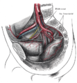
 The peritoneum of the male pelvis. | |
| Details | |
| Identifiers | |
| Latin | plica umbilicalis lateralis; plica epigastrica |
| TA98 | A10.1.02.434 |
| TA2 | 3796 |
| FMA | 16537 |
| Anatomical terminology | |
The lateral umbilical fold is an elevation (on either side of the body) of the peritoneum lining the inner/posterior surface of the lower anterior abdominal wall formed by the underlying inferior epigastric artery and inferior epigastric vein which the peritoneum covers. Superiorly, the lateral umbilical fold ends where the vessels reach and enter the rectus sheath[1] at the arcuate line of rectus sheath; in spite of the name, the lateral umbilical folds do not extend as far superiorly as the umbilicus.[2] Inferiorly, it extends to just medial to the deep inguinal ring.[citation needed]
Each lateral umbilical fold is situated lateral to the ipsilateral medial umbilical fold. Unlike the median and medial umbilical folds, the contents of the lateral umbilical fold remain functional after birth.[2]
Clinical significance
[edit]The lateral umbilical fold is an important reference site with regards to hernia classification. A direct hernia occurs medial to the lateral umbilical fold, whereas an indirect hernia originates lateral to the fold. This latter case is due to the placement of the opening of the deep inguinal ring in the space lateral to the lateral umbilical fold, which allows the passage of the ductus deferens, testicular artery, and other components of the spermatic cord in men, or the round ligament of the uterus in women.[citation needed]
Additional images
[edit]-
The arteries of the pelvis.
See also
[edit]References
[edit]- ^ Standring, Susan (2020). Gray's Anatomy: The Anatomical Basis of Clinical Practice (42th ed.). New York. p. 1156. ISBN 978-0-7020-7707-4. OCLC 1201341621.
{{cite book}}: CS1 maint: location missing publisher (link) - ^ a b Sinnatamby, Chummy (2011). Last's Anatomy (12th ed.). Elsevier Australia. p. 234. ISBN 978-0-7295-3752-0.
![]() This article incorporates text in the public domain from page 1152 of the 20th edition of Gray's Anatomy (1918)
This article incorporates text in the public domain from page 1152 of the 20th edition of Gray's Anatomy (1918)
External links
[edit]- Lateral umbilical fold
- Anatomy figure: 36:03-09 at Human Anatomy Online, SUNY Downstate Medical Center - "Internal surface of the anterior abdominal wall."
- Anatomy image:7384 at the SUNY Downstate Medical Center

