Vestibular membrane: Difference between revisions
{{Short description}}. Sectioning as per WP:ANATMOS. →Structure: Reference for separating scalae. |
|||
| Line 1: | Line 1: | ||
{{Short description|Membrane in the cochlea in the inner ear}} |
|||
| ⚫ | |||
{{Infobox anatomy |
{{Infobox anatomy |
||
| Name = Vestibular membrane |
| Name = Vestibular membrane |
||
| Line 10: | Line 12: | ||
| Location = [[Cochlea]] of the [[inner ear]] |
| Location = [[Cochlea]] of the [[inner ear]] |
||
| Pronunciation = {{IPAc-en|lang|ˈ|r|aɪ|s|n|ər}} |
| Pronunciation = {{IPAc-en|lang|ˈ|r|aɪ|s|n|ər}} |
||
}} |
|||
| ⚫ | |||
| ⚫ | The '''vestibular membrane''', '''vestibular wall''' or '''Reissner's membrane''', is a [[diaphragm (acoustics)|membrane]] inside the [[cochlea]] of the [[inner ear]]. It separates the [[cochlear duct]] from the [[vestibular duct]]. Together with the [[basilar membrane]], it creates a compartment in the cochlea filled with [[endolymph]], which is important for the function of the spiral [[organ of Corti]]. It |
||
| ⚫ | The '''vestibular membrane''', '''vestibular wall''' or '''Reissner's membrane''', is a [[diaphragm (acoustics)|membrane]] inside the [[cochlea]] of the [[inner ear]]. It separates the [[cochlear duct]] from the [[vestibular duct]]. Together with the [[basilar membrane]], it creates a compartment in the cochlea filled with [[endolymph]], which is important for the function of the spiral [[organ of Corti]]. It allows nutrients to travel from the [[perilymph]] to the [[endolymph]] of the [[membranous labyrinth]]. It is named after the German [[anatomist]] [[Ernst Reissner]]. |
||
== Structure == |
|||
The vestibular membrane separates the [[cochlear duct]] (scala media) from the [[vestibular duct]] (scala vestibuli).<ref>{{Cite book|last=Javel|first=Eric|url=https://www.sciencedirect.com/science/article/pii/B0122268709005293|title=Encyclopedia of the Neurological Sciences|publisher=[[Academic Press]]|year=2003|isbn=978-0-12-226870-0|pages=305-311|language=en|chapter=Auditory System, Peripheral|doi=10.1016/B0-12-226870-9/00529-3}}</ref> |
|||
=== Microanatomy === |
|||
[[Histologically]], the membrane is composed of two layers of flattened [[epithelium]], separated by a [[basal lamina]]. Its structure suggests that its function is transport of fluid and [[electrolytes]].{{uncited|date=April 2020}} |
[[Histologically]], the membrane is composed of two layers of flattened [[epithelium]], separated by a [[basal lamina]]. Its structure suggests that its function is transport of fluid and [[electrolytes]].{{uncited|date=April 2020}} |
||
== Function == |
|||
| ⚫ | |||
Together with the [[basilar membrane]], the vestibular membrane creates a compartment in the cochlea filled with [[endolymph]]. This is important for the function of the spiral [[organ of Corti]]. It primarily functions as a [[diffusion]] barrier, allowing nutrients to travel from the [[perilymph]] to the [[endolymph]] of the [[membranous labyrinth]]. |
|||
== |
== History == |
||
| ⚫ | |||
| ⚫ | |||
== Additional images == |
|||
| ⚫ | |||
File:Gray929.png|Floor of cochlear duct. |
File:Gray929.png|Floor of cochlear duct. |
||
File:Gray930.png|Spiral limbus and basilar membrane. |
File:Gray930.png|Spiral limbus and basilar membrane. |
||
</gallery> |
</gallery> |
||
== |
== References == |
||
<references /> |
|||
== External links == |
|||
* {{KansasHistology|eye_ear|ear03}} |
* {{KansasHistology|eye_ear|ear03}} |
||
* {{UIUCHistologySubject|76}} |
* {{UIUCHistologySubject|76}} |
||
Revision as of 15:41, 11 December 2021
This article includes a list of references, related reading, or external links, but its sources remain unclear because it lacks inline citations. (June 2015) |
| Vestibular membrane | |
|---|---|
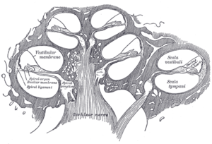
 Cross-section of the cochlea showing the position of the vestibular membrane. | |
 Cross-section of the cochlea at higher magnification showing the membrane (here labelled "Reissner's membrane") | |
| Details | |
| Pronunciation | English: /ˈraɪsnər/ |
| Location | Cochlea of the inner ear |
| Identifiers | |
| Latin | membrana vestibularis ductus cochlearis |
| Anatomical terminology | |
The vestibular membrane, vestibular wall or Reissner's membrane, is a membrane inside the cochlea of the inner ear. It separates the cochlear duct from the vestibular duct. Together with the basilar membrane, it creates a compartment in the cochlea filled with endolymph, which is important for the function of the spiral organ of Corti. It allows nutrients to travel from the perilymph to the endolymph of the membranous labyrinth. It is named after the German anatomist Ernst Reissner.
Structure
The vestibular membrane separates the cochlear duct (scala media) from the vestibular duct (scala vestibuli).[1]
Microanatomy
Histologically, the membrane is composed of two layers of flattened epithelium, separated by a basal lamina. Its structure suggests that its function is transport of fluid and electrolytes.[citation needed]
Function
Together with the basilar membrane, the vestibular membrane creates a compartment in the cochlea filled with endolymph. This is important for the function of the spiral organ of Corti. It primarily functions as a diffusion barrier, allowing nutrients to travel from the perilymph to the endolymph of the membranous labyrinth.
History
The vestibular membrane is also known as Reissner's membrane. This alternative name is named after German anatomist Ernst Reissner (1824-1878).
Additional images
-
Floor of cochlear duct.
-
Spiral limbus and basilar membrane.
References
- ^ Javel, Eric (2003). "Auditory System, Peripheral". Encyclopedia of the Neurological Sciences. Academic Press. pp. 305–311. doi:10.1016/B0-12-226870-9/00529-3. ISBN 978-0-12-226870-0.

