Sartorius muscle: Difference between revisions
modified caption to be more accurate (see Quadriceps article) |
No edit summary |
||
| Line 50: | Line 50: | ||
==Common Injuries of the Sartorius Muscle== |
==Common Injuries of the Sartorius Muscle== |
||
Overextension of the [[hip]] may cause a [[ |
Overextension of the [[hip]] may cause a [[strain]] of the muscle at its attachment point (the [[iliac crest]]). |
||
==Additional images== |
==Additional images== |
||
Revision as of 20:18, 19 April 2007
This December 2006 may be confusing or unclear to readers. |
| Sartorius muscle | |
|---|---|
 Muscles of lower extremity. (Rectus femoris removed to reveal the vastus intermedius.) | |
| Details | |
| Origin | superior to the anterior superior iliac spine |
| Insertion | medial side of the upper tibia in the pes anserinus |
| Artery | femoral artery |
| Nerve | femoral nerve |
| Actions | Flexion of knee, Flexion of leg |
| Identifiers | |
| Latin | musculus sartorius |
| TA98 | A04.7.02.015 |
| TA2 | 2610 |
| FMA | 22353 |
| Anatomical terms of muscle | |
The sartorius muscle is a long thin muscle that runs down the length of the thigh. It is the longest muscle in the human body. Its upper portion forms the lateral border of the femoral triangle.
Origin and insertion
The sartorius muscle arises by tendinous fibers from the anterior superior iliac spine, running obliquely across the upper and anterior part of the thigh in an inferomedial direction.
It descends as far as the medial side of the knee, passing behind the medial condyle of the femur to end in a tendon.
This tendon curves anteriorly to join the tendons of the gracilis and semitendinous muscles which together form the pes anserinus, finally inserting into the proximal part of the tibia on the medial surface of its body.
Etymology
The name sartorius is the Latin word for "sartorial" (i.e. "to do with tailoring", in turn from sartor i.e. "tailor", in turn from sartus i.e. "patched" or "repaired", in turn from sarcio i.e. "to patch", "to repair").
This name was chosen in reference to the cross-legged position in which tailors once sat.
Action
The action of sartorius is to cross the legs, by flexion of the knee, and flexion and lateral rotation of the hip. Sartorius does not have a very strong action.
Innervation
Situated in the anterior fascial compartment of the thigh, sartorius is innervated via branches of the femoral nerve.
Variations
Slips of origin from the outer end of the inguinal ligament, the notch of the ilium, the ilio-pectineal line or the pubis occur.
The muscle may be split into two parts, and one part may be inserted into the fascia lata, the femur, the ligament of the patella or the tendon of the Semitendinosus.
The tendon of insertion may end in the fascia lata, the capsule of the knee-joint, or the fascia of the leg.
The muscle may be absent[citation needed].
Common Injuries of the Sartorius Muscle
Overextension of the hip may cause a strain of the muscle at its attachment point (the iliac crest).
Additional images
-
Bones of the right leg. Anterior surface.
-
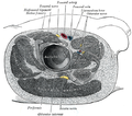
Structures surrounding right hip-joint.
-
Muscles of the iliac and anterior femoral regions.
-
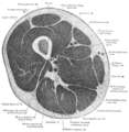
Cross-section through the middle of the thigh.
-
Muscles of the gluteal and posterior femoral regions.
-
Femoral sheath laid open to show its three compartments.
-
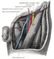
The left femoral triangle.
-
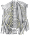
The lumbar plexus and its branches.
-
Front and medial aspect of right thigh.
External links
- . GPnotebook https://www.gpnotebook.co.uk/simplepage.cfm?ID=208339022.
{{cite web}}: Missing or empty|title=(help) - Template:MuscleLoyola
- Anatomy photo:14:st-0407 at the SUNY Downstate Medical Center
- Cross section image: pembody/body15a—Plastination Laboratory at the Medical University of Vienna
- Cross section image: pelvis/pelvis-e12-15—Plastination Laboratory at the Medical University of Vienna
![]() This article incorporates text in the public domain from page 470 of the 20th edition of Gray's Anatomy (1918)
This article incorporates text in the public domain from page 470 of the 20th edition of Gray's Anatomy (1918)


