Ejaculatory duct: Difference between revisions
Appearance
Content deleted Content added
→External links: stub sorting |
They do not 'cause the reflex action of ejaculation'; formation; length; redid opening line; link to Excretory duct of seminal gland |
||
| Line 13: | Line 13: | ||
MeshNumber = A05.360.444.251 | |
MeshNumber = A05.360.444.251 | |
||
}} |
}} |
||
The '''Ejaculatory ducts''' are |
The '''Ejaculatory ducts''' are paired structures in male [[anatomy]], about 2 cm in length. |
||
Each ejaculatory duct is formed by the union of the [[vas deferens]] with the [[Excretory duct of seminal gland|duct of the seminal vesicle]]. They pass through the [[prostate]], and empty into the [[urethra]] at the [[Colliculus seminalis]]. During ejaculation, [[semen]] passes through the ducts and exits the body via the [[penis]]. |
|||
Surgery to correct [[Benign Prostatic Hyperplasia]] (BPH) may destroy these ducts resulting in[[ retrograde ejaculation]]. |
Surgery to correct [[Benign Prostatic Hyperplasia]] (BPH) may destroy these ducts resulting in[[ retrograde ejaculation]]. |
||
Revision as of 14:56, 9 October 2007
| Ejaculatory duct | |
|---|---|
| File:Male anatomy.png Male Anatomy | |
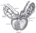
 Vesiculæ seminales and ampullæ of ductus deferentes, seen from the front. The anterior walls of the left ampulla, left seminal vesicle, and prostatic urethra have been cut away. | |
| Details | |
| Identifiers | |
| Latin | ductus ejaculatorii |
| MeSH | D004543 |
| TA98 | A09.3.07.001 |
| TA2 | 3636 |
| FMA | 19325 |
| Anatomical terminology | |
The Ejaculatory ducts are paired structures in male anatomy, about 2 cm in length.
Each ejaculatory duct is formed by the union of the vas deferens with the duct of the seminal vesicle. They pass through the prostate, and empty into the urethra at the Colliculus seminalis. During ejaculation, semen passes through the ducts and exits the body via the penis.
Surgery to correct Benign Prostatic Hyperplasia (BPH) may destroy these ducts resulting inretrograde ejaculation.
Additional images
-
Lobes of prostate
-
Vertical section of bladder, penis, and urethra.
-
Prostate with seminal vesicles and seminal ducts, viewed from in front and above.
-
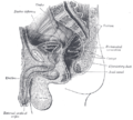
Median sagittal section of male pelvis.
See also
External links
- Anatomy figure: 44:03-15 at Human Anatomy Online, SUNY Downstate Medical Center - "Lateral (A) and posterior (B) views of the bladder and associated structures."


