Glans penis: Difference between revisions
| [pending revision] | [pending revision] |
m Revert previous revision by 155.245.112.207 |
ScrotalRaphe (talk | contribs) →Additional images: Photo of ventral view of galsn penis. |
||
| Line 49: | Line 49: | ||
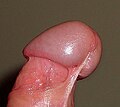
Image:Penisfrenulum.jpg|The glans penis of an uncircumcised male. |
Image:Penisfrenulum.jpg|The glans penis of an uncircumcised male. |
||
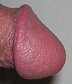
Image:Side_View_Glans.JPG|Side view circumcised glans penis |
Image:Side_View_Glans.JPG|Side view circumcised glans penis |
||
Image:Anteriorglanspenis.jpg|An [[anterior]] |
Image:Anteriorglanspenis.jpg|An [[anterior]] View of a flaccid uncircumcised glans penis |
||
Image:VentralViewGlansPenis.JPG|Ventral view of gpans penis |
|||
</gallery> |
</gallery> |
||
Revision as of 07:24, 7 February 2008
| male sexual organs | |
|---|---|
| File:Male anatomy.png 1. Testicles 2. Epididymis 3. Corpus cavernosa 4. Foreskin 5. Frenulum 6. Urethral opening 7. 8. Corpus spongiosum 9. Penis 10. Scrotum | |
| Details | |
| Artery | Urethral artery |
| Identifiers | |
| Latin | GraySubject = 262 |
| TA98 | A09.4.01.007 |
| TA2 | 3668 |
| FMA | 18247 |
| Anatomical terminology | |
The glans penis (or simply glans) is the sensitive tip of the penis. It is also commonly referred to as the "head" of the penis. Slang terms include "helmet" and "bell end". When the penis is flaccid it is wholly or partially covered by the foreskin, except in men who have been circumcised.
Medical considerations
The meatus (opening) of the urethra is at the tip of the glans penis. In circumcised infants who wear diapers, the meatal area of the glans penis is without the protection of the foreskin and at slight risk of meatitis, meatal ulceration, and meatal stenosis.[1]
The epithelium of the glans penis is mucocutaneous tissue.[2] Birley et al. report that excessive washing with soap may dry the mucous membrane that covers the glans penis and cause non-specific dermatitis.[3]
Inflammation of the glans penis is known as balanitis. It occurs in 3–11% of males, and up to 35% of diabetic males. It has many causes, including irritation, or infection with a wide variety of pathogens. Careful identification of the cause with the aid of patient history, physical examination, swabs and cultures, and biopsy are essential in order to determine the proper treatment.[4]
Anatomical details
The glans penis is the expanded cap of the corpus spongiosum. It is moulded on the rounded ends of the Corpora cavernosa penis, extending farther on their upper than on their lower surfaces. At the summit of the glans is the slit-like vertical external urethral orifice. The circumference of the base of the glans forms a rounded projecting border, the corona glandis, overhanging a deep retroglandular sulcus (the coronal sulcus), behind which is the neck of the penis. The proportional size of the glans penis can vary greatly.
The foreskin maintains the mucosa in a moist environment.[5] In males who have been circumcised, but have not undergone restoration, the glans is permanently exposed and dry. Szabo and Short found that the glans of the circumcised penis does not develop a thicker keratinization layer.[6] Studies have suggested that the glans is equally sensitive in circumcised and uncircumcised males.[7] [8]
Halata & Munger (1986) report that the density of genital corpuscles is greatest in the corona glandis,[9] while Yang & Bradley (1998) report that their study "showed no areas in the glans to be more densely innervated than others."[10]
Halata & Spathe (1997) reported that "the glans penis contains a predominance of free nerve endings, numerous genital end bulbs and rarely Pacinian and Ruffinian corpuscles. Merkel nerve endings and Meissner corpuscles are not present."[2]
Yang & Bradley argue that "The distinct pattern of innervation of the glans emphasizes the role of the glans as a sensory structure".[10]
An anatomycal variant of glans is showed: Hirsuties papillaris genitalis
Additional images
-
Diagram of the arteries of the penis.
-
Penis
-
The glans penis of an uncircumcised male.
-
Side view circumcised glans penis
-
An anterior View of a flaccid uncircumcised glans penis
-
Ventral view of gpans penis
See also
References
- ^ Freud, Paul (1947). "The ulcerated urethral meatus in male children". The Journal of Pediatrics. 31 (2): 131–41. doi:10.1016/S0022-3476(47)80098-8. Retrieved 2006-07-07.
{{cite journal}}: Unknown parameter|month=ignored (help) - ^ a b Halata, Zdenek (1997). "Sensory innervation of the human penis". Advances in experimental medicine and biology (424): 265–6. PMID 9361804. Retrieved 2006-07-07.
{{cite journal}}: Unknown parameter|coauthors=ignored (|author=suggested) (help) - ^ Birley, H. D. (1993). "Clinical features and management of recurrent balanitis; association with atopy and genital washing". Genitourinary Medicine. 69 (5): 400–3. PMID 8244363.
{{cite journal}}: Unknown parameter|coauthors=ignored (|author=suggested) (help); Unknown parameter|month=ignored (help); line feed character in|coauthors=at position 67 (help) - ^ Edwards, Sarah (1996). "Balanitis and balanoposthitis: a review". Genitourinary Medicine. 72 (3): 155–9. PMID 8707315.
{{cite journal}}: Unknown parameter|month=ignored (help) - ^ Prakash, Satya (1982). "Sub-Preputial Wetness--Its Nature". Annals Of National Medical Science (India). 18 (3): 109–112.
{{cite journal}}: Unknown parameter|coauthors=ignored (|author=suggested) (help); Unknown parameter|month=ignored (help) - ^ Szabo, Robert (2000). "How does male circumcision protect against HIV infection?". British Medical Journal. 320 (7249): 1592–4. PMID 10845974. Retrieved 2006-07-07.
{{cite journal}}: Unknown parameter|coauthors=ignored (|author=suggested) (help); Unknown parameter|month=ignored (help) - ^ Masters, William H. (1966). Human Sexual Response. Boston: Little, Brown & Co. pp. 189–91. ISBN 0-316-54987-8.
{{cite book}}: Unknown parameter|coauthors=ignored (|author=suggested) (help) (excerpt accessible here) - ^ Bleustein, Clifford B. (2005). "Effect of neonatal circumcision on penile neurologic sensation". Urology. 65 (4): 773–7. doi:10.1016/j.urology.2004.11.007. PMID 15833526.
{{cite journal}}: Unknown parameter|coauthors=ignored (|author=suggested) (help); Unknown parameter|month=ignored (help) - ^ Halata, Zdenek (1986). "The neuroanatomical basis for the protopathic sensibility of the human glans penis". Brain Research. 371 (2): 205–30. doi:10.1016/0006-8993(86)90357-4. PMID 3697758.
{{cite journal}}: Unknown parameter|coauthors=ignored (|author=suggested) (help); Unknown parameter|month=ignored (help) - ^ a b Yang, C. C. (1998). "Neuroanatomy of the penile portion of the human dorsal nerve of the penis". British Journal of Urology. 82 (1): 109–13. doi:10.1046/j.1464-410x.1998.00669.x. PMID 9698671.
{{cite journal}}: Unknown parameter|coauthors=ignored (|author=suggested) (help); Unknown parameter|month=ignored (help)
External links
- Anatomy photo:42:07-0102 at the SUNY Downstate Medical Center - "The Male Perineum and the Penis: The Corpus Spongiosum and Corpora Cavernosa"
- Anatomy photo:44:06-0101 at the SUNY Downstate Medical Center - "The Male Pelvis: The Urethra"


