Central nervous system: Difference between revisions
No edit summary |
Revert to revision 612459636 dated 2014-06-11 05:59:52 by 117.239.68.146 using popups |
||
| Line 1: | Line 1: | ||
{{redirect|CNS}} |
|||
{{Infobox anatomy |
{{Infobox anatomy |
||
| Name = Central nervous system |
| Name = Central nervous system |
||
Revision as of 11:28, 12 June 2014
| Central nervous system | |
|---|---|
 Schematic diagram showing the central nervous system in pink, peripheral in yellow. | |
| Details | |
| Identifiers | |
| Latin | Systema nervosum centrale pars centralis systematis nervosi[1] |
| MeSH | D002490 |
| TA98 | A14.1.00.001 |
| TA2 | 5364 |
| FMA | 55675 |
| Anatomical terminology | |
The central nervous system (CNS) is the part of the nervous system consisting of the brain and spinal cord. The peripheral nervous system (or PNS), is composed of nerves leading to and from the CNS, often through junctions known as ganglia.[citation needed]
The central nervous system is so named because it integrates information it receives from, and coordinates and influences the activity of, all parts of the bodies of bilaterally symmetric animals—that is, all multicellular animals except sponges and radially symmetric animals such as jellyfish, and it contains the majority of the nervous system.[citation needed] Arguably many consider the retina[2] and the optic nerve (2nd cranial nerve),[3][4] as well as the olfactory nerves (1st) and olfactory epithelium[5] as parts of the CNS, synapsing directly on brain tissue without intermediate ganglia. Following this classification the olfactory epithelium is the only central nervous tissue in direct contact with the environment, which opens up for therapeutic treatments. [5] The CNS is contained within the dorsal cavity, with the brain in the cranial cavity and the spinal cord in the spinal cavity. In vertebrates, the brain is protected by the skull, while the spinal cord is protected by the vertebrae, both enclosed in the meninges.[6]
Structure
The central nervous system consists of the two major structures, the brain and spinal cord. The brain is encased in the skull, and protected by the cranium.[7] The spinal cord is continuous with the brain and lies caudaly to the brain,[8] and is protected by the vertebra.[7] The spinal cord reaches from the base of the skull, continues through[7] or starting below[9] the foramen magnum,[7] and terminates roughly level with the first or second lumbar vertebra,[8][9] occupying the upper sections of the vertebral canal.[4]
White and gray matter

Microscopically, there are differences between the neurons and tissue of the central nervous system and the peripheral nervous system.[citation needed] The central nervous system is divided in white and gray matter.[8] This can also be seen macroscopically on brain tissue. The white matter constitutes of axons and oligodendrocytes, while the gray matter chiefly constitutes of neurons. Both tissues include a number of glial cells (although the white matter contains more), which are often referred to as supporting cells of the central nervous system. Different forms of glial cells have different functions, some acting almost as scaffolding for neuroblasts to climb during neurogenesis such as bergmann glia, while others such as microglia are a specialized form of macrophage, involved in the immune system of the brain as well as the clearance of various metabolites from the brain tissue.[4] Astrocytes may be involved with both clearance of metabolites as well as transport of fuel and various beneficial substances to neurons from the capillaries of the brain. Upon CNS injury astrocytes will proliferate, causing gliosis, a form of neuronal scar tissue, lacking in functional neurons.[4]
The brain (cerebrum as well as midbrain & hindbrain) consists of a cortex, composed of neuron-bodies constituting gray matter, while internally there is more white matter which form tracts and commissures. Apart from cortical gray matter there is also subcortical gray making up a large number of different nuclei.[8]
Spinal cord
From and to the spinal cord are projections of the peripheral nervous system in the form of spinal nerves (sometimes segmental nerves[7]). The nerves connect the spinal cord with skin, joints, muscles etc. and allow for the transmission of efferent motor as well as afferent sensory signals and stimuli.[8] This allows for voluntary and involuntary motions of muscles, as well as the perception of senses. All in all 31 spinal nerves project from the brain stem,[8] some forming plexa as they branch out, such as the; brachial plexa, sacral plexa etc.[7] Each spinal nerve will carry both sensory and motor signals, but the nerves synapse at different regions of the spinal cord, either from the periphery to sensory relay neurons which relay the information to the CNS or from the CNS to motor neurons, which relay the information out.[8]


The spinal cord relays information up to the brain through spinal tracts through the "final common pathway" [8] to the thalamus and ultimately to the cortex. Not all information is relayed to the cortex, and does not reach our immediate consciousness, but is instead transmitted only to the thalamus which sorts and adapts accordingly. This in turn may explain why we are not constantly aware of all aspects of our surroundings.[citation needed]
-
Schematic image showing the locations of a few tracts of the spinal cord.
-
Reflexes may also occur without engaging more than one neuron of the central nervous system as in the below example of a short reflex.
Cranial nerves
Apart from the spinal cord, there are also peripheral nerves of the PNS that synapse through intermediaries or ganglia directly on the CNS. These 12 nerves exist in the head and neck region and are called cranial nerves. Cranial nerves bring information to the CNS to and from the face, as well as to certain muscles (such as the trapezius muscle, which is innervated by accessory nerves[7] as well as certain cervical spinal nerves).[7]
Two pairs of cranial nerves; the olfactory nerves and the optic nerves[2] are often considered structures of the central nervous system. This is because they do not synapse first on peripheral ganglia, but directly on central nervous neurons. The olfactory epithelium is significant in that it consists of central nervous tissue expressed in direct contact to the environment, allowing for administration of certain pharmaceuticals and drugs. [5]
Brain
Rostrally to the spinal cord lies the brain.[8] The brain makes up the largest portion of the central nervous system, and is often the main structure referred to when speaking of the nervous system. The brain is the major functional unit of the central nervous system. While the spinal cord has certain processing ability such as that of spinal locomotion and can process reflexes, the brain is the major processing unit of the nervous system.[citation needed]
Brain stem
The brain stem consists of the medulla, the pons and the midbrain. The medulla can be referred to as an extension of the spinal cord, and its organization and functional properties are similar to those of the spinal cord.[8] The tracts passing from the spinal cord to the brain pass through here.[8]
Regulatory functions of the medulla nuclei include control of the blood pressure and breathing. Other nuclei are involved in balance, taste, hearing and control of muscles of the face and neck.[8]
The next structure rostral to the medulla is the pons, which lies on the ventral anterior side of the brain stem. Nuclei in the pons include pontine nuclei which work with the cerebellum and transmit information between the cerebellum and the cerebral cortex.[8] In the dorsal posterior pons lie nuclei that have to do with breathing, sleep and taste.[8]
The midbrain (or mesencephalon) is situated above and rostral to the pons, and includes nuclei linking distinct parts of the motor system, among others the cerebellum, the basal ganglia and both cerebral hemispheres. Additionally parts of the visual and auditory systems are located in the mid brain, including control of automatic eye movements.[8]
The brain stem at large provides entry and exit to the brain for a number of pathways for motor and autonomic control of the face and neck through cranial nerves,[8] and autonomic control of the organs is mediated by the tenth cranial (vagus) nerve.[4] A large portion of the brain stem is involved in such autonomic control of the body. Such functions may engage the heart, blood vessels, pupillae, among others.[8]
The brain stem also hold the reticular formation, a group of nuclei involved in both arousal and alertness.[8]
Cerebellum
The cerebellum lies posteriorly or dorsally and rostrally to the pons. The cerebellum is composed of several dividing fissures and lobes. Its function includes posture and coordination of movements of eyes, limbs as well as that of the head. Further it is involved in motion that has been learned and perfected though practice, and will adapt to new learned movements.[8] Despite its previous classification as a motor structure, the cerebellum also displays connections to areas of the cerebral cortex involved in language as well as cognitive functions. These connections have been recently shown through anatomical studies, such as fMRI and PET.[8]
The body of the cerebellum holds more neurons than any other structure of the brain including that of the larger cerebrum (or cerebral hemispheres), but is also more extensively understood than other structures of the brain, and includes fewer types of different neurons.[8] It handles and processes sensory stimuli, motor information as well as balance information from the vestibular organ.[8]
Diencephalon
The two structures of the diecephalon worth noting are the thalamus and the hypothalamus. The thalamus acts as a linkage between incoming pathways from the peripheral nervous system as well as the optical nerve (though it does not receive input from the olfactory nerve) to the cerebral hemispheres. Previously it was considered only a "relay station", but it is engaged in the sorting of information that will reach cerebral hemispheres (neocortex).[8]
Apart from its function of sorting information from the periphery, the thalamus also connects the cerebellum and basal ganglia with the cerebrum. In common with the aforementioned reticular system the thalamus is involved in wakefullness and consciousness, such as though the SCN.[8]
The hypothalamus engages in functions of a number of primitive emotions or feelings such as hunger, thirst and maternal bonding. This is regulated partly through control of secretion of hormones from the pituitary gland. Additionally the hypothalamus plays a role in motivation and many other behaviors of the individual.[8]
Cerebrum
The cerebrum of cerebral hemispheres make up the largest portion of the human brain. Various structures combine forming the cerebral hemispheres, among others, the cortex, basal ganglia, amygdala and hippocampus. The hemispheres together control a large portion of the functions of the human brain such as emotion, memory, perception and motor functions. Apart from this the cerebral hemispheres stand for the cognitive capabilities of the brain.[8]
Connecting each of the hemispheres is the corpus callosum as well as several additional commissures.[8] One of the most important parts of the cerebral hemispheres is the cortex, which is made up of gray matter covering the surface of the brain. Functionally, the cerebral cortex is involved in planning and carrying out of everyday tasks.[8]
The hippocampus is involved in storage of memories, the amygdala plays a role in perception and communication of emotion, while the basal ganglia play a major role in the coordination of voluntary movement.[8]
Difference from the peripheral nervous system

This differentiates the central nervous system from the peripheral nervous system, which consists of neurons, axons and Schwann cells. Schwann cells and oligodendrocytes have similar functions in the central and peripheral nervous system respectively. Both act to add myelin sheaths to the axons, which acts as a form of insulation allowing for better and faster proliferation of electrical signals along the nerves. Axons in the central nervous system are often very short (barely a few millimeters) and do not need the same degree of isolation as peripheral nerves do. Some peripheral nerves can be over 1m in length, such as the nerves to the big toe. To ensure signals move at sufficient speed, myelination is needed.
The way in which the Schwann cells and oligodendrocytes myelinate nerves differ. A Schwann cell usually myelinates a single axon, completely surrounding it. Sometimes the it may myelinate many axons, especially when in areas of short ones.[7] Oligodendrocytes usually myelinate several axons. They do this by sending out thin projections of their cell membrane which envelop and enclose the axon.
Development
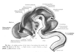
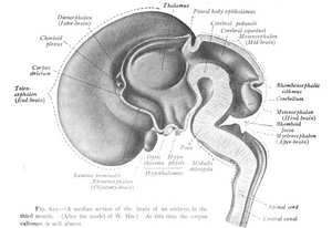
During early development of the vertebrate embryo, a longitudinal groove on the neural plate gradually deepens and the ridges on either side of the groove (the neural folds) become elevated, and ultimately meet, transforming the groove into a closed tube, the ectodermal wall of which forms the rudiment of the nervous system. This tube initially differentiates into three vesicles (pockets): the prosencephalon at the front, the mesencephalon, and, between the mesencephalon and the spinal cord, the rhombencephalon. (By six weeks in the human embryo) the prosencephalon then divides further into the telencephalon and diencephalon; and the rhombencephalon divides into the metencephalon and myelencephalon.
As a vertebrate grows, these vesicles differentiate further still. The telencephalon differentiates into, among other things, the striatum, the hippocampus and the neocortex, and its cavity becomes the first and second ventricles. Diencephalon elaborations include the subthalamus, hypothalamus, thalamus and epithalamus, and its cavity forms the third ventricle. The tectum, pretectum, cerebral peduncle and other structures develop out of the mesencephalon, and its cavity grows into the mesencephalic duct (cerebral aqueduct). The metencephalon becomes, among other things, the pons and the cerebellum, the myelencephalon forms the medulla oblongata, and their cavities develop into the fourth ventricle.
-
Development of the neural tube
Evolution
Planarians, members of the phylum Platyhelminthes (flatworms), have the simplest, clearly defined delineation of a nervous system into a central nervous system (CNS) and a peripheral nervous system (PNS).[10][11] Their primitive brains, consisting of two fused anterior ganglia, and longitudinal nerve cords form the CNS; the laterally projecting nerves form the PNS. A molecular study found that more than 95% of the 116 genes involved in the nervous system of planarians, which includes genes related to the CNS, also exist in humans.[12] Like planarians, vertebrates have a distinct CNS and PNS, though more complex than those of planarians.
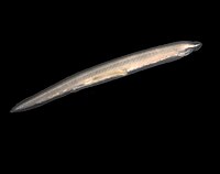
The CNS of chordates differs from that of other animals in being placed dorsally in the body, above the gut and notochord/spine.[13] The basic pattern of the CNS is highly conserved throughout the different species of vertebrates and during evolution. The major trend that can be observed is towards a progressive telencephalisation: the telencephalon of reptiles is only an appendix to the large olfactory bulb, while in mammals it makes up most of the volume of the CNS. In the human brain, the telencephalon covers most of the diencephalon and the mesencephalon. Indeed, the allometric study of brain size among different species shows a striking continuity from rats to whales, and allows us to complete the knowledge about the evolution of the CNS obtained through cranial endocasts.
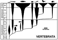
Mammals – which appear in the fossil record after the first fishes, amphibians, and reptiles – are the only vertebrates to possess the evolutionarily recent, outermost part of the cerebral cortex known as the neocortex.[14] The neocortex of monotremes (the duck-billed platypus and several species of spiny anteaters) and of marsupials (such as kangaroos, koalas, opossums, wombats, and Tasmanian devils) lack the convolutions – gyri and sulci – found in the neocortex of most placental mammals (eutherians).[15] Within placental mammals, the size and complexity of the neocortex increased over time. The area of the neocortex of mice is only about 1/100 that of monkeys, and that of monkeys is only about 1/10 that of humans.[14] In addition, rats lack convolutions in their neocortex (possibly also because rats are small mammals), whereas cats have a moderate degree of convolutions, and humans have quite extensive convolutions.[14] Extreme convolution of the neocortex is found in dolphins, possibly related to their complex echolocation.
Diseases
There are many central nervous system diseases and conditions, including infections of the central nervous system such as encephalitis and poliomyelitis, early-onset neurological disorders including ADHD and autism, late-onset neurodegenerative diseases such as Alzheimer's disease, Parkinson's disease, and essential tremor, autoimmune and inflammatory diseases such as multiple sclerosis and acute disseminated encephalomyelitis, genetic disorders such as Krabbe's disease and Huntington's disease, as well as amyotrophic lateral sclerosis and adrenoleukodystrophy. Lastly, cancers of the central nervous system can cause severe illness and, when malignant, can have very high mortality rates.
Specialty professional organizations recommend that neurological imaging of the brain be done only to answer a specific clinical question and not as routine screening.[16]
References
- ^ Farlex Partner Medical Dictionary, Farlex 2012.
- ^ a b Purves, Dale (2000). Neuroscience, Second Edition. Sunderland, MA: Sinauer Associates. ISBN 9780878937424.
- ^ "Medical Subject Headings (MeSH): Optic Nerve". National Library of Medicine. Retrieved 28 September 2013.
- ^ a b c d e Estomih Mtui, M.J. Turlough FitzGerald, Gregory Gruener. Clinical neuroanatomy and neuroscience (6th ed. ed.). Edinburgh: Saunders. p. 38. ISBN 978-0-7020-3738-2.
{{cite book}}:|edition=has extra text (help)CS1 maint: multiple names: authors list (link) - ^ a b c Gizurarson S (2012). "Anatomical and histological factors affecting intranasal drug and vaccine delivery". Current Drug Delivery. 9 (6): 566–582. doi:10.2174/156720112803529828. PMC 3480721. PMID 22788696.
- ^ Maton, Anthea; Jean Hopkins; Charles William McLaughlin; Susan Johnson; Maryanna Quon Warner; David LaHart; Jill D. Wright (1993). Human Biology and Health. Englewood Cliffs, New Jersey, USA: Prentice Hall. pp. 132–144. ISBN 0-13-981176-1.
- ^ a b c d e f g h i Arthur F. Dalley, Keith L. Moore, Anne M.R. Agur (2010). Clinically oriented anatomy (6th ed., [International ed.]. ed.). Philadelphia [etc.]: Lippincott Williams & Wilkins, Wolters Kluwer. pp. 48–55, 464, 700, 822, 824, 1075. ISBN 978-1-60547-652-0.
{{cite book}}: CS1 maint: multiple names: authors list (link) - ^ a b c d e f g h i j k l m n o p q r s t u v w x y z aa ab ac Principles of neural science (5. ed. ed.). Appleton and Lange: McGraw Hill. 2012. pp. 338–343. ISBN 978-0-07-139011-8.
{{cite book}}:|edition=has extra text (help);|first=has generic name (help);|first=missing|last=(help) - ^ a b Huijzen, R. Nieuwenhuys, J. Voogd, C. van (2008). The human central nervous system (4th ed. ed.). Berlin: Springer. p. 3. ISBN 978-3-540-34686-9.
{{cite book}}:|edition=has extra text (help)CS1 maint: multiple names: authors list (link) - ^ Hickman, Jr., Cleveland P.; Larry S. Roberts; Susan L. Keen; Allan Larson; Helen L'Anson; David J. Eisenhour (2008). Integrated Princinples of Zoology: Fourteenth Edition. New York, NY, USA: McGraw-Hill Higher Education. p. 733. ISBN 978-0-07-297004-3.
- ^ Campbell, Neil A.; Jane B. Reece; Lisa A. Urry; Michael L. Cain; Steven A. Wasserman; Peter V. Minorsky; Robert B. Jackson (2008). Biology: Eighth Edition. San Francisco, CA, USA: Pearson / Benjamin Cummings. p. 1065. ISBN 978-0-8053-6844-4.
- ^ Mineta K, Nakazawa M, Cebria F, Ikeo K, Agata K, Gojobori T (2003). "Origin and evolutionary process of the CNS elucidated by comparative genomics analysis of planarian ESTs". PNAS. 100 (13): 7666–7671. doi:10.1073/pnas.1332513100. PMC 164645. PMID 12802012.
{{cite journal}}: CS1 maint: multiple names: authors list (link) - ^ Romer, A.S. (1949): The Vertebrate Body. W.B. Saunders, Philadelphia. (2nd ed. 1955; 3rd ed. 1962; 4th ed. 1970)
- ^ a b c Bear, Mark F.; Barry W. Connors; Michael A. Paradiso (2007). Neuroscience: Exploring the Brain: Third Edition. Philadelphia, PA, USA: Lippincott Williams & Wilkins. pp. 196–199. ISBN 978-0-7817-6003-4. Cite error: The named reference "MarkFBear" was defined multiple times with different content (see the help page).
- ^ Kent, George C.; Robert K. Carr (2001). Comparative Anatomy of the Vertebrates: Ninth Edition. New York, NY, USA: McGraw-Hill Higher Education. p. 409. ISBN 0-07-303869-5.
- ^ American College of Radiology; American Society of Neuroradiology (2010). "ACR-ASNR practice guideline for the performance of computed tomography (CT) of the brain". Agency for Healthcare Research and Quality. Reston, VA, USA: American College of Radiology. Retrieved 9 September 2012Template:Inconsistent citations
{{cite journal}}: CS1 maint: postscript (link)
External links
- Overview of the Central Nervous System, Neuroscience Online (electronic neuroscience textbook)
- High-Resolution Cytoarchitectural Primate Brain Atlases
- Explaining the human nervous system.
- The Department of Neuroscience at Wikiversity


