Ischioanal fossa: Difference between revisions
Appearance
Content deleted Content added
Bot: Removing Commons:File:Gray405.png (en). It was deleted on Commons by INeverCry (Per Commons:Commons:Deletion requests/penis). |
Courcelles (talk | contribs) m Reverted edits by Filedelinkerbot (talk) to last version by Werieth |
||
| Line 4: | Line 4: | ||
| GraySubject = 120 |
| GraySubject = 120 |
||
| GrayPage = 425 |
| GrayPage = 425 |
||
| Image = |
| Image = Gray405.png |
||
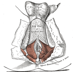
| Caption = The [[perineum]]. The integument and superficial layer of [[superficial fascia]] reflected. (Ischiorectal fossa labeled at bottom left.) |
|||
| Caption = |
|||
| Image2 = Gray1077.png |
| Image2 = Gray1077.png |
||
| Caption2 = The posterior aspect of the rectum exposed by removing the lower part of the sacrum and the coccyx. (Ischiorectal fossa labeled at bottom right.) |
| Caption2 = The posterior aspect of the rectum exposed by removing the lower part of the sacrum and the coccyx. (Ischiorectal fossa labeled at bottom right.) |
||
Revision as of 03:27, 6 September 2014
| Ischioanal fossa | |
|---|---|
 The perineum. The integument and superficial layer of superficial fascia reflected. (Ischiorectal fossa labeled at bottom left.) | |
 The posterior aspect of the rectum exposed by removing the lower part of the sacrum and the coccyx. (Ischiorectal fossa labeled at bottom right.) | |
| Details | |
| Identifiers | |
| Latin | fossa ischioanalis |
| TA98 | A09.5.04.001 |
| TA2 | 2446 |
| FMA | 22059 |
| Anatomical terminology | |
The ischioanal fossa (formerly called ischiorectal fossa) is the fat-filled space located lateral to the anal canal and inferior to the pelvic diaphragm. It is somewhat prismatic in shape, with its base directed to the surface of the perineum, and its apex at the line of meeting of the obturator and anal fasciae.
Boundaries
It has the following boundaries:[1]
| ANTERIOR * fascia of Colles covering the Transversus perinei superficialis * inferior fascia of the urogenital diaphragm |
||
| LATERAL * tuberosity of the ischium * Obturator internus muscle * obturator fascia |
SUPERIOR: * Levator ani INFERIOR: |
MEDIAL: * Levator ani * Sphincter ani externus muscle * anal fascia |
| POSTERIOR * Gluteus maximus * sacrotuberous ligament |
Contents
The contents include:
- Inside Alcock's canal, on the lateral wall
- Outside Alcock's canal, crossing the space transversely
- inferior rectal artery
- inferior rectal veins
- inferior anal nerves
- fatty tissue across which numerous fibrous bands extend from side to side
See also
References
- ^ analtrianglesection - Ischiorectal fossa is colored yellow
Additional images
-
Coronal section of anterior part of pelvis, through the pubic arch. Seen from in front.
-
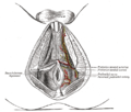
The superficial branches of the internal pudendal artery.
External links
- Anatomy image: apmalefrontal4-8 at the College of Medicine at SUNY Upstate Medical University
- Anatomy photo:41:04-0103 at the SUNY Downstate Medical Center - "The Female Perineum: The Ischioanal Fossa"
- Anatomy image:9246 at the SUNY Downstate Medical Center
- Cross section image: pelvis/pelvis-e12-15—Plastination Laboratory at the Medical University of Vienna
- perineum at The Anatomy Lesson by Wesley Norman (Georgetown University) (analtriangle2, femaleugtrianglesectionfascia)
- Diagram at Emory University
![]() This article incorporates text in the public domain from page 425 of the 20th edition of Gray's Anatomy (1918)
This article incorporates text in the public domain from page 425 of the 20th edition of Gray's Anatomy (1918)


