User:ThunderPop/sandbox: Difference between revisions
ThunderPop (talk | contribs) Rearrangement, advantages & limitations, history |
ThunderPop (talk | contribs) |
||
| Line 20: | Line 20: | ||
Samples spotted on a SELDI surface are typically analyzed using time-of-flight mass spectrometry. An irradiating laser ionizes peptides from crystals of the sample/matrix mixture. The ions are then briefly accelerated through an [[electric potential]] and travel down a field-free flight tube where they are separated by their [[velocity]] differences. The [[mass-to-charge ratio]] of each ion can be determined from the length of the tube, the [[kinetic energy]] given to ions by the electric field, and the velocity of the ions in the tube. The velocity of the ions is inversely proportional to the square root of the mass-to-charge ratio of the ion; ions with low mass-to-charge ratios are detected earlier than ions with high mass-to-charge ratios.<ref>{{Cite book|url=http://doi.wiley.com/10.1002/0470118490|title=Fundamentals of Contemporary Mass Spectrometry - Dass - Wiley Online Library|last=Dass|first=Chhabil|doi=10.1002/0470118490}}</ref> |
Samples spotted on a SELDI surface are typically analyzed using time-of-flight mass spectrometry. An irradiating laser ionizes peptides from crystals of the sample/matrix mixture. The ions are then briefly accelerated through an [[electric potential]] and travel down a field-free flight tube where they are separated by their [[velocity]] differences. The [[mass-to-charge ratio]] of each ion can be determined from the length of the tube, the [[kinetic energy]] given to ions by the electric field, and the velocity of the ions in the tube. The velocity of the ions is inversely proportional to the square root of the mass-to-charge ratio of the ion; ions with low mass-to-charge ratios are detected earlier than ions with high mass-to-charge ratios.<ref>{{Cite book|url=http://doi.wiley.com/10.1002/0470118490|title=Fundamentals of Contemporary Mass Spectrometry - Dass - Wiley Online Library|last=Dass|first=Chhabil|doi=10.1002/0470118490}}</ref> |
||
Binding to the SELDI surface acts as a separation step, and as a result, the proteins bound to the surface are easier to analyze. The surface is composed primarily of materials with a variety of physico-chemical characteristics, metal ions, or anion or cation exchangers. Common surfaces include CM10 (weak cation [[Ion exchange|exchange]]), H50 (hydrophobic surface, similar to C<sub>6</sub>-C<sub>12</sub> [[reverse phase chromatography]]), IMAC30 (metal-binding surface), and Q10 (strong anion exchange). SELDI surfaces can also be functionalized to study DNA-protein binding, antibody-antigen assays, and receptor-ligand interactions.<ref name=":1" /> |
Binding to the SELDI surface acts as a solid-phase chromatographic separation step, and as a result, the proteins bound to the surface are easier to analyze. The surface is composed primarily of materials with a variety of physico-chemical characteristics, metal ions, or anion or cation exchangers. Common surfaces include CM10 (weak cation [[Ion exchange|exchange]]), H50 (hydrophobic surface, similar to C<sub>6</sub>-C<sub>12</sub> [[reverse phase chromatography]]), IMAC30 (metal-binding surface), and Q10 (strong anion exchange). SELDI surfaces can also be functionalized to study DNA-protein binding, antibody-antigen assays, and receptor-ligand interactions.<ref name=":1" /> |
||
== History == |
== History == |
||
| Line 31: | Line 31: | ||
== Applications == |
== Applications == |
||
SELDI-TOF-MS is optimal for analyzing low molecular weight proteins (<20 kDa) in a variety of biological materials, such as tissue samples, blood, urine, and serum. This technique is often used in combination with [[Western blot|immunoblotting]] and [[immunohistochemistry]] to aid in the detection of biomarkers for diseases, and has also been applied to the diagnosis of cancer and neurological disorders.<ref>{{Cite journal|last=Gloerich|first=Jolein|last2=Wevers|first2=Ron A.|last3=Smeitink|first3=Jan A. M.|last4=Engelen|first4=Baziel G. van|last5=Heuvel|first5=Lambert P. van den|date=2006-12-07|title=Proteomics Approaches to Study Genetic and Metabolic Disorders|url=http://pubs.acs.org/doi/abs/10.1021/pr060487w|journal=Journal of Proteome Research|language=en|volume=6|issue=2|pages=506–512|doi=10.1021/pr060487w}}</ref> |
SELDI-TOF-MS is optimal for analyzing low molecular weight proteins (<20 kDa) in a variety of biological materials, such as tissue samples, blood, urine, and serum. This technique is often used in combination with [[Western blot|immunoblotting]] and [[immunohistochemistry]] to aid in the detection of biomarkers for diseases, and has also been applied to the diagnosis of cancer and neurological disorders.<ref name=":5">{{Cite journal|last=Gloerich|first=Jolein|last2=Wevers|first2=Ron A.|last3=Smeitink|first3=Jan A. M.|last4=Engelen|first4=Baziel G. van|last5=Heuvel|first5=Lambert P. van den|date=2006-12-07|title=Proteomics Approaches to Study Genetic and Metabolic Disorders|url=http://pubs.acs.org/doi/abs/10.1021/pr060487w|journal=Journal of Proteome Research|language=en|volume=6|issue=2|pages=506–512|doi=10.1021/pr060487w}}</ref> SELDI technology is most widely used to compare serum samples from healthy and diseased patients. Serum studies allow for a minimally invasive approach to disease monitoring in patients and are useful in the early detection and diagnosis of diseases and neurological disorders, such as [[amyotrophic lateral sclerosis]] (ALS).<ref name=":5" /><ref name=":6" /> |
||
== Advantages & Limitations == |
== Advantages & Limitations == |
||
A major advantage of the SELDI process is that sample components that interfere with other analytical tools, including salts, detergents, buffers, or other compounds, are removed before analysis with mass spectrometry. Only the proteins that are specifically bound to the spot surfaces are analyzed, reducing the overall complexity of the sample. As a result, there is an increased probability of detecting analytes that are present in lower concentrations.<ref>{{Cite journal|last=Seibert|first=Volker|last2=Wiesner|first2=Andreas|last3=Buschmann|first3=Thomas|last4=Meuer|first4=Jörn|date=2004-04-30|title=Surface-enhanced laser desorption ionization time-of-flight mass spectrometry (SELDI TOF-MS) and ProteinChip® technology in proteomics research|url=http://www.sciencedirect.com/science/article/pii/S0344033804000263|journal=Pathology - Research and Practice|series=Proteomics in Pathology, Research and Practice|volume=200|issue=2|pages=83–94|doi=10.1016/j.prp.2004.01.010}}</ref> In proteomics, the biomarker discovery, identification, and validation steps can all be done on the SELDI surface.<ref name=":0" /> |
A major advantage of the SELDI process is that sample components that interfere with other analytical tools, including salts, detergents, buffers, or other compounds, are removed before analysis with mass spectrometry. Only the proteins that are specifically bound to the spot surfaces are analyzed, reducing the overall complexity of the sample. As a result, there is an increased probability of detecting analytes that are present in lower concentrations.<ref name=":6">{{Cite journal|last=Seibert|first=Volker|last2=Wiesner|first2=Andreas|last3=Buschmann|first3=Thomas|last4=Meuer|first4=Jörn|date=2004-04-30|title=Surface-enhanced laser desorption ionization time-of-flight mass spectrometry (SELDI TOF-MS) and ProteinChip® technology in proteomics research|url=http://www.sciencedirect.com/science/article/pii/S0344033804000263|journal=Pathology - Research and Practice|series=Proteomics in Pathology, Research and Practice|volume=200|issue=2|pages=83–94|doi=10.1016/j.prp.2004.01.010}}</ref> In proteomics, the biomarker discovery, identification, and validation steps can all be done on the SELDI surface.<ref name=":0" /> |
||
Contrastingly, SELDI is often criticized for its [[reproducibility]] due to differences in the mass spectra obtained when using different batches of chip surfaces. There also exists a potential for sample bias, as nonspecific absorption matrices favor the binding of analytes with higher abundancies in the sample at the expense of less abundant analytes.<ref name=":1" /> |
Contrastingly, SELDI is often criticized for its [[reproducibility]] due to differences in the mass spectra obtained when using different batches of chip surfaces. There also exists a potential for sample bias, as nonspecific absorption matrices favor the binding of analytes with higher abundancies in the sample at the expense of less abundant analytes.<ref name=":1" /> Additionally, the baseline signal in the spectra varies and noise from the matrix is maximal below 2000 Da, with Ciphergen Biosystems suggesting to ignore spectral peaks below 2000 Da.<ref>{{Cite journal|last=Henderson|first=N.A.|last2=Steele|first2=R.J.C.|title=SELDI-TOF proteomic analysis and cancer detection|url=http://linkinghub.elsevier.com/retrieve/pii/S1479666X05800484|journal=The Surgeon|volume=3|issue=6|pages=383–390|doi=10.1016/s1479-666x(05)80048-4}}</ref> |
||
==See also== |
==See also== |
||
Revision as of 02:01, 28 March 2016
| Acronym | SELDI |
|---|---|
| Analytes | Biomolecules |
| Other techniques | |
| Related | Matrix-assisted laser desorption/ionization Soft laser desorption Surface-assisted laser desorption/ionization |
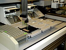
Surface-enhanced laser desorption/ionization (SELDI) is a soft ionization method in mass spectrometry (MS) used for the analysis of protein mixtures. It is a variation of matrix-assisted laser desorption/ionization (MALDI) and incorporates surface-enhanced neat desorption (SEND),surface-enhanced affinity-capture (SEAC), and surface-enhanced photolabile attachment and release (SEPAR) mass spectrometry.[1] [2] SELDI is typically used with time-of-flight (TOF) mass spectrometers and is used to detect proteins in tissue samples, blood, urine, or other clinical samples. Comparison of protein levels between patients with and without a disease can be used for biomarker discovery.[3][4]
Sample Preparation & Instrumentation

SELDI can be seen as a combination of solid-phase chromatography and TOF-MS. The sample is spotted onto a modified chip surface, where each spot on the surface allows for the specific binding of proteins from the sample. Contaminants and nonspecifically bound proteins are then washed away. After washing the spotted sample, an energy absorbing matrix, such as sinapinic acid (SPA) or α-Cyano-4-hydroxycinnamic acid (CHCA), is applied to the surface and allowed to crystallize with the sample.[1][5] Alternatively, the matrix can be attached to the sample surface by covalent modification or adsorption before the sample is spotted.[2] The sample is then irradiated by a pulsed laser, causing ablation and desorption of the sample and matrix.[1][5]
Samples spotted on a SELDI surface are typically analyzed using time-of-flight mass spectrometry. An irradiating laser ionizes peptides from crystals of the sample/matrix mixture. The ions are then briefly accelerated through an electric potential and travel down a field-free flight tube where they are separated by their velocity differences. The mass-to-charge ratio of each ion can be determined from the length of the tube, the kinetic energy given to ions by the electric field, and the velocity of the ions in the tube. The velocity of the ions is inversely proportional to the square root of the mass-to-charge ratio of the ion; ions with low mass-to-charge ratios are detected earlier than ions with high mass-to-charge ratios.[6]
Binding to the SELDI surface acts as a solid-phase chromatographic separation step, and as a result, the proteins bound to the surface are easier to analyze. The surface is composed primarily of materials with a variety of physico-chemical characteristics, metal ions, or anion or cation exchangers. Common surfaces include CM10 (weak cation exchange), H50 (hydrophobic surface, similar to C6-C12 reverse phase chromatography), IMAC30 (metal-binding surface), and Q10 (strong anion exchange). SELDI surfaces can also be functionalized to study DNA-protein binding, antibody-antigen assays, and receptor-ligand interactions.[5]
History

SELDI technology was developed by T. William Hutchens and Tai-Tung Yip at Baylor College of Medicine in 1993.[7] Hutchens and Yip attached single-stranded DNA to agarose beads and used the beads to capture lactoferrin, an iron-binding glycoprotein, from preterm infant urine. The beads were incubated in the sample and then removed, washed, and analyzed with a MALDI-MS probe tip. This research led to the idea that MALDI surfaces could be derivatized with SEAC devices; the technique was later described by Hutchens and Yip in 1998.[1][7]
The SELDI process is a combination of SEND, SEAC, and SEPAR technology. With SEND, analytes can be desorbed and ionized without adding a matrix; the matrix is incorporated into the sample surface.In SEAC, the sample surface is derivatized to bind the analyte of interest for analysis with laser desorption/ionization mass spectrometry (LDI-MS).[1][2][8] SEPAR is a hybrid of SEND and SEAC; the derivatized sample surface also acts as an energy absorbing matrix for ionization.[2]
SELDI technology was first commercialized by Ciphergen Biosystems in 1997 as the ProteinChip system, and is now produced and marketed by Bio-Rad Laboratories.[8]
Applications
SELDI-TOF-MS is optimal for analyzing low molecular weight proteins (<20 kDa) in a variety of biological materials, such as tissue samples, blood, urine, and serum. This technique is often used in combination with immunoblotting and immunohistochemistry to aid in the detection of biomarkers for diseases, and has also been applied to the diagnosis of cancer and neurological disorders.[9] SELDI technology is most widely used to compare serum samples from healthy and diseased patients. Serum studies allow for a minimally invasive approach to disease monitoring in patients and are useful in the early detection and diagnosis of diseases and neurological disorders, such as amyotrophic lateral sclerosis (ALS).[9][10]
Advantages & Limitations
A major advantage of the SELDI process is that sample components that interfere with other analytical tools, including salts, detergents, buffers, or other compounds, are removed before analysis with mass spectrometry. Only the proteins that are specifically bound to the spot surfaces are analyzed, reducing the overall complexity of the sample. As a result, there is an increased probability of detecting analytes that are present in lower concentrations.[10] In proteomics, the biomarker discovery, identification, and validation steps can all be done on the SELDI surface.[1]
Contrastingly, SELDI is often criticized for its reproducibility due to differences in the mass spectra obtained when using different batches of chip surfaces. There also exists a potential for sample bias, as nonspecific absorption matrices favor the binding of analytes with higher abundancies in the sample at the expense of less abundant analytes.[5] Additionally, the baseline signal in the spectra varies and noise from the matrix is maximal below 2000 Da, with Ciphergen Biosystems suggesting to ignore spectral peaks below 2000 Da.[11]
See also
References
- ^ a b c d e f Tang N, Tornatore P, Weinberger SR (2004). "Current developments in SELDI affinity technology". Mass spectrometry reviews. 23 (1): 34–44. doi:10.1002/mas.10066. PMID 14625891.
{{cite journal}}: CS1 maint: multiple names: authors list (link) - ^ a b c d Merchant, Maggie; Weinberger, Scot R. (2000-04-01). "Recent advancements in surface-enhanced laser desorption/ionization-time of flight-mass spectrometry". ELECTROPHORESIS. 21 (6): 1164–1177. doi:10.1002/(SICI)1522-2683(20000401)21:63.0.CO;2-0. ISSN 1522-2683.
- ^ Li J, Zhang Z, Rosenzweig J, Wang YY, Chan DW (2002). "Proteomics and bioinformatics approaches for identification of serum biomarkers to detect breast cancer". Clin. Chem. 48 (8): 1296–304. PMID 12142387.
{{cite journal}}: CS1 maint: multiple names: authors list (link) - ^ Jr GW, Cazares LH, Leung SM, Nasim S, Adam BL, Yip TT, Schellhammer PF, Gong L, Vlahou A (1999). "Proteinchip(R) surface enhanced laser desorption/ionization (SELDI) mass spectrometry: a novel protein biochip technology for detection of prostate cancer biomarkers in complex protein mixtures". Prostate Cancer and Prostatic Diseases. 2 (5/6): 264–276. doi:10.1038/sj.pcan.4500384. PMID 12497173.
{{cite journal}}: CS1 maint: multiple names: authors list (link) - ^ a b c d Muthu, Manikandan; Vimala, A.; Mendoza, Ordetta Hanna; Gopal, Judy (2016-02-01). "Tracing the voyage of SELDI-TOF MS in cancer biomarker discovery and its current depreciation trend – need for resurrection?". TrAC Trends in Analytical Chemistry. 76: 95–101. doi:10.1016/j.trac.2015.10.004.
- ^ Dass, Chhabil. Fundamentals of Contemporary Mass Spectrometry - Dass - Wiley Online Library. doi:10.1002/0470118490.
- ^ a b Hutchens TW and Yip TT. "New desorption strategies for the mass spectrometric analysis of macromolecules." Rapid Commun Mass Spectrom 7: 576-580 (1993). [1]
- ^ a b Lomas, Lee O.; Weinberger, Scot R. (2008-01-01). Surface-Enhanced Laser Desorption/Ionization (SELDI) Technology. John Wiley & Sons, Ltd. doi:10.1002/9780470061565.hbb128. ISBN 9780470061565.
- ^ a b Gloerich, Jolein; Wevers, Ron A.; Smeitink, Jan A. M.; Engelen, Baziel G. van; Heuvel, Lambert P. van den (2006-12-07). "Proteomics Approaches to Study Genetic and Metabolic Disorders". Journal of Proteome Research. 6 (2): 506–512. doi:10.1021/pr060487w.
- ^ a b Seibert, Volker; Wiesner, Andreas; Buschmann, Thomas; Meuer, Jörn (2004-04-30). "Surface-enhanced laser desorption ionization time-of-flight mass spectrometry (SELDI TOF-MS) and ProteinChip® technology in proteomics research". Pathology - Research and Practice. Proteomics in Pathology, Research and Practice. 200 (2): 83–94. doi:10.1016/j.prp.2004.01.010.
- ^ Henderson, N.A.; Steele, R.J.C. "SELDI-TOF proteomic analysis and cancer detection". The Surgeon. 3 (6): 383–390. doi:10.1016/s1479-666x(05)80048-4.
