Vestibular membrane: Difference between revisions
m Dating maintenance tags: {{Uncited}} |
m svg |
||
| Line 5: | Line 5: | ||
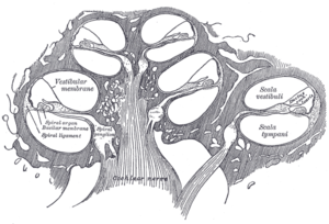
| Caption = Cross-section of the [[cochlea]] showing the position of the vestibular membrane. |
| Caption = Cross-section of the [[cochlea]] showing the position of the vestibular membrane. |
||
| Width = 300 |
| Width = 300 |
||
| Image2 = Cochlea-crosssection. |
| Image2 = Cochlea-crosssection.svg |
||
| Caption2 = Cross-section of the cochlea at higher magnification showing the membrane (here labelled "Reissner's membrane") |
| Caption2 = Cross-section of the cochlea at higher magnification showing the membrane (here labelled "Reissner's membrane") |
||
| System = |
| System = |
||
Revision as of 16:09, 27 June 2020
| Vestibular membrane | |
|---|---|
 Cross-section of the cochlea showing the position of the vestibular membrane. | |
 Cross-section of the cochlea at higher magnification showing the membrane (here labelled "Reissner's membrane") | |
| Details | |
| Pronunciation | English: /ˈraɪsnər/ |
| Location | Cochlea of the inner ear |
| Identifiers | |
| Latin | membrana vestibularis ductus cochlearis |
| Anatomical terminology | |
This article includes a list of references, related reading, or external links, but its sources remain unclear because it lacks inline citations. (June 2015) |
The vestibular membrane, vestibular wall or Reissner's membrane, is a membrane inside the cochlea of the inner ear. It separates the cochlear duct from the vestibular duct. Together with the basilar membrane it creates a compartment in the cochlea filled with endolymph, which is important for the function of the spiral organ of Corti. It primarily functions as a diffusion barrier, allowing nutrients to travel from the perilymph to the endolymph of the membranous labyrinth.
Histologically, the membrane is composed of two layers of flattened epithelium, separated by a basal lamina. Its structure suggests that its function is transport of fluid and electrolytes.[citation needed]
Reissner's membrane is named after German anatomist Ernst Reissner (1824-1878).
Additional images
-
Floor of cochlear duct.
-
Spiral limbus and basilar membrane.

