Trapezius
This December 2006 may be confusing or unclear to readers. |
| Trapezius | |
|---|---|
 Trapezius. | |
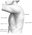
 Muscles connecting the upper extremity to the vertebral column. (Trapezius visible at upper right.) | |
| Details | |
| Origin | arises, down the midline, from the external occipital protuberance, the nuchal ligament, the medial part of the superior nuchal line, and the spinous processes of the vertebrae C7-T12 |
| Insertion | at the shoulders, into the lateral third of the clavicle, the acromion process, and into the spine of the scapula |
| Nerve | major nerve supply is the cranial nerve XI. Cervical nerves C3 and C4 receive information about pain in this muscle |
| Actions | retraction of scapula |
| Antagonist | Serratus anterior muscle |
| Identifiers | |
| Latin | musculus trapezius |
| TA98 | A04.3.01.001 |
| TA2 | 2226 |
| FMA | 9626 |
| Anatomical terms of muscle | |
In human anatomy, the trapezius is a large superficial muscle on a person's back.
Actions
Because the fibers run in different directions, it has a variety of actions, including:
- scapular elevation (shrugging up or lifting the shoulders),
- scapular adduction (drawing the shoulder blades)
- scapular depression (pulling the shoulder blades down)
Different fibers control different actions:
- The superior (upper) fibers elevate the scapula.
- the middle fibers retract it.
- The inferior (lower) fibers depress it.
- When the superior and inferior fibers act together they superiorly (upwardly) rotate the scapula.
How to injure:
Tumbling head first over mountain bike handles and landing on the back of the head can pull the Trapezius muscle. The resulting pull can:
- create an inflamation of the muscle tissue
- the inflamation can constrict the nerves to the arm leaving a tingling or numbness
- cause a stiff neck
Nomenclature
Trapezius gets its name from its trapezium-like shape; the corners being the neck, the two shoulders, and the thoracic vertebra, T12.
The word "Spinotrapezius", when applied to humans, refers to the trapezius, although it not commonly used in modern texts. In other mammals, it refers to a portion of the analogous muscle, it gives you a sort of a Arch on/near your neck. See trapezius muscles (cat) for more details.
Exercise
The upper portion of the trapezius can be developed by elevating the shoulders. Common exercises for this movement are shoulder shrugs and upright rows. Middle fibers are developed by pulling shoulder blades together. Best exercises for this movement are rowing exercises and deadlifts. Lower part can be developed by drawing the shoulder blades downward while keeping arms almost straight and stiff. This can be done in a pull-down station for example.
A person can feel the muscles of the trapezius become active by holding a weight in front of them in one hand, and with the other, touching the area between the shoulder and the neck. It is common for non-experienced gym users to focus mostly to the upper portion of the muscle, and thus forgotting important middle part and creating muscle imbalances which can heavily affect posture and compromise shoulder health.
Anatomical details
It arises from the external occipital protuberance and the medial third of the superior nuchal line of the occipital bone, from the ligamentum nuchae, the spinous process of the seventh cervical, and the spinous processes of all the thoracic vertebræ, and from the corresponding portion of the supraspinal ligament.
From this origine:
- the superior fibers proceed downward and lateralward. They are inserted into the posterior border of the lateral third of the clavicle.
- the inferior fibers proceed upward and lateralward. They converge near the scapula, and end in an aponeurosis, which glides over the smooth triangular surface on the medial end of the spine, to be inserted into a tubercle at the apex of this smooth triangular surface.
- the middle fibers proceed horizontally. They are inserted into the medial margin of the acromion, and into the superior lip of the posterior border of the spine of the scapula.
At its occipital origin, the Trapezius is connected to the bone by a thin fibrous lamina, firmly adherent to the skin.
At the middle it is connected to the spinous processes by a broad semi-elliptical aponeurosis, which reaches from the sixth cervical to the third thoracic vertebræ, and forms, with that of the opposite muscle, a tendinous ellipse.
The rest of the muscle arises by numerous short tendinous fibers.
The two Trapezius muscles together resemble a trapezium, or diamond-shaped quadrangle: two angles corresponding to the shoulders; a third to the occipital protuberance; and the fourth to the spinous process of the twelfth thoracic vertebra.
Variations
The attachments to the dorsal vertebrae are often reduced and the lower ones are often wanting; the occipital attachment is often wanting; separation between cervical and dorsal portions is frequent.
Extensive deficiencies and complete absence occur.
The clavicular insertion of this muscle varies in extent; it sometimes reaches as far as the middle of the clavicle, and occasionally may blend with the posterior edge of the Sternocleidomastoideus, or overlap it.
Additional images
-
Muscles of head and neck.
-
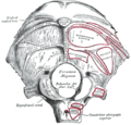
Occipital bone. Outer surface.
-
Left clavicle. Superior surface.
-
Left scapula. Dorsal surface.
-
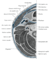
Section of the neck at about the level of the sixth cervical vertebra.
-
Muscles of the neck. Anterior view.
-
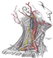
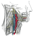
Superficial dissection of the right side of the neck, showing the carotid and subclavian arteries.
-
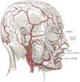
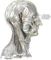
The arteries of the face and scalp.
-
The triangles of the neck.
-
The nerves of the scalp, face, and side of neck.
-
Hypoglossal nerve, cervical plexus, and their branches.
-
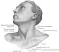
Anterolateral view of head and neck.
-
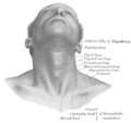
Front view of neck.
-
Surface anatomy of the back.
-
The left side of the thorax.
External links
- Template:MuscleLoyola
- . GPnotebook https://www.gpnotebook.co.uk/simplepage.cfm?ID=724893774.
{{cite web}}: Missing or empty|title=(help) - Illustration at preventdisease.com
- Template:Exrx
- Photograph at mgccc.cc.ms.us
![]() This article incorporates text in the public domain from page 432 of the 20th edition of Gray's Anatomy (1918)
This article incorporates text in the public domain from page 432 of the 20th edition of Gray's Anatomy (1918)


