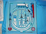
Central venous catheter


In medicine, a central venous catheter ("central line", "CVC", "central venous line" or "central venous access catheter") is a catheter placed into a large vein in the neck (internal jugular vein or external jugular vein), chest (subclavian vein) or groin (femoral vein). It is used to administer medication or fluids, obtain blood tests (specifically the "mixed venous oxygen saturation"), and directly obtain cardiovascular measurements such as the central venous pressure.
Types
There are several types of central venous catheters:[1]
Tunneled catheter
This type of catheter is inserted into a vein at one location (neck, chest or groin), and tunneled under the skin to a separate exit site, where it emerges from underneath the skin. It is held in place by a Dacron cuff, just underneath the skin at the exit site. The exit site is typically located in the chest, making the access ports less visible than if they were to directly protrude from the neck. Passing the catheter under the skin helps to prevent infection and provides stability.
Implanted port

A port is similar to a tunneled catheter but is left entirely under the skin. Medicines are injected through the skin into the catheter. Some implanted ports contain a small reservoir that can be refilled in the same way. After being filled, the reservoir slowly releases the medicine into the bloodstream. An implanted port is less obvious than a tunneled catheter and requires very little daily care. It has less impact on a person's activities than a PICC line or a tunneled catheter. Surgically implanted infusion port placed below the clavicle (infraclavicular fossa), catheter threaded into the right atrium through large vein. Accessed via non-coring "Huber" needle through the skin. May need to use topical anesthetic prior to accessing port. Used for medications, chemotherapy, TPN, and blood. Easy to maintain for home-based therapy.
PICC line
A peripherally inserted central catheter, or PICC line (pronounced "pick"), is a central venous catheter inserted into a vein in the arm rather than a vein in the neck or chest.
Technical description

Dependent on its use, the catheter is monoluminal, biluminal or triluminal, dependent on the actual number of tubes or lumens (1, 2 and 3 respectively). Some catheters have 4 or 5 lumens, depending on the reason for their use.
The catheter is usually held in place by a suture or staple and an occlusive dressing. Regular flushing with saline or a heparin-containing solution keeps the line patent and prevents thrombosis. Certain lines are impregnated with antibiotics, silver-containing substances (specifically silver sulfadiazine) and/or chlorhexidine to reduce infection risk.
Specific types of long-term central lines are the Hickman catheters, which require clamps to make sure the valve is closed, and Groshong catheters, which have a valve that opens as fluid is withdrawn or infused and remains closed when not in use. Hickman and Groshong lines need more specific measures to prevent infection. Hence, they are inserted into the jugular vein but then tunneled under the skin to maximize the distance a pathogen would need to travel to enter the bloodstream. Hickman lines also have a "cuff" under the skin, again to prevent bacterial migration.[citation needed]
Indications and uses
Indications for the use of central lines include:[2]
- Monitoring of the central venous pressure (CVP) in acutely ill patients to quantify fluid balance
- Long-term Intravenous antibiotics
- Long-term Parenteral nutrition especially in chronically ill patients
- Long-term pain medications
- Chemotherapy
- Drugs that are prone to cause phlebitis in peripheral veins (caustic), such as:
- Calcium chloride
- Chemotherapy
- Hypertonic saline
- Potassium chloride
- Amiodarone
- Plasmapheresis
- Dialysis
- Frequent blood draws
- Frequent or persistent requirement for intravenous access
- Need for intravenous therapy when peripheral venous access is impossible
Central venous catheters usually remain in place for a longer period of time, especially when the reason for their use is longstanding (such as total parenteral nutrition in a chronically ill patient). For such indications, a Hickman line, a PICC line or a portacath may be considered because of their smaller infection risk. Sterile technique is highly important here, as a line may serve as a porte d'entrée (place of entry) for pathogenic organisms, and the line itself may become infected with organisms such as Staphylococcus aureus and coagulase-negative Staphylococci.[citation needed]
Insertion


The skin is cleaned, and local anesthetic applied if required. The location of the vein is then identified by landmarks or with the use of a small ultrasound device. A hollow needle is advanced through the skin until blood is aspirated; the color of the blood and the rate of its flow help distinguish it from arterial blood (suggesting that an artery has been accidentally punctured).[citation needed]
The Seldinger technique is then employed to insert the line. This means that a blunt guidewire is passed through the needle, and the needle is then removed. A dilating device may be passed over the guidewire to slightly enlarge the tract, and the central line itself is then passed over the guidewire, which is then removed. All the lumens of the line are aspirated (to ensure that they are all positioned inside the vein) and flushed.[citation needed]
For jugular and subclavian lines, a chest X-ray is typically performed to ensure the line is positioned inside the superior vena cava and, in the case of insertion through the subclavian vein, that there is no resultant pneumothorax.
The technique for placement of a central venous catheter is demonstrated in the video[3]. A central venous catheter can be placed under ultrasound guidance as in the video[4].
Complications
Central line insertion may cause a number of complications. The benefit expected from their use therefore needs to outweigh the risk of those complications.
Pneumothorax
Pneumothorax (for central lines placed in the chest); the incidence is thought to be higher with subclavian vein catheterization. In catheterization of the internal jugular vein, the risk of pneumothorax can be minimized by the use of ultrasound guidance. For experienced clinicians, the incidence of pneumothorax is about 1%. Some official bodies, e.g. the National Institute for Health and Clinical Excellence (UK), recommend the routine use of ultrasonography to minimize complications.[5]
Infection
All catheters can introduce bacteria into the bloodstream, but CVCs are known for occasionally causing Staphylococcus aureus and Staphylococcus epidermidis sepsis. Infection risks were initially thought to be less in jugular lines, but this only seems to be the case if the patient is obese.[6]
If a patient with a central line develops signs of infection, blood cultures are taken from both the catheter and from a vein elsewhere in the body. If the culture from the central line grows bacteria much earlier (>2 hours) than the other site, the line is the likely source of the infection. Quantitative blood culture is even more accurate, but this is not widely available.[7]
Generally, antibiotics are used, and occasionally the catheter will have to be removed. In the case of bacteremia from Staphylococcus aureus, removing the catheter without administering antibiotics is not adequate as 38% of such patients may still develop endocarditis.[8]
In a clinical practice guideline, the American Centers for Disease Control and Prevention recommends against routine culturing of central venous lines upon their removal.[9] The guideline makes a number of further recommendations to prevent line infections.[9]
To prevent infection, stringent cleaning of the catheter insertion site is advised. Povidone-iodine solution is often used for such cleaning, but chlorhexidine appears to be twice as good as iodine.[10] Routine replacement of lines makes no difference in preventing infection.[11]
Other complications
Rarely, small amounts of air are sucked into the vein as a result of the negative Intra-thoracic pressure.[citation needed] If these air bubbles obstruct blood vessels, this is known as an air embolism.
Hemorrhage (bleeding) and formation of a hematoma (bruise) is slightly more common in jugular venous lines than in others.[6]
Arrhythmias may occur during the insertion process when the wire comes in contact with the endocardium. It typically resolved when the wire is pulled back.[citation needed]
References
- ^ Central Venous Catheters - Topic Overview from WebMD
- ^ Central Venous Catheter Placement - Department of Surgery, Baylor College of Medicine, Texas, Houston
- ^ http://www.dailymotion.com/video/x2gm9v_central-venous-catheter-placement-p_tech
- ^ http://in.youtube.com/watch?v=Ahz1SPKTiBU
- ^ National Institute for Health and Clinical Excellence (2002). "Technology appraisal: the clinical effectiveness and cost effectiveness of ultrasonic locating devices for the placement of central venous lines". Retrieved 2008-06-01.
{{cite web}}: Unknown parameter|month=ignored (help) - ^ a b Parienti JJ, Thirion M, Mégarbane B; et al. (2008). "Femoral vs jugular venous catheterization and risk of nosocomial events in adults requiring acute renal replacement therapy: a randomized controlled trial". JAMA. 299 (20): 2413–22. doi:10.1001/jama.299.20.2413. PMID 18505951.
{{cite journal}}: Explicit use of et al. in:|author=(help); Unknown parameter|month=ignored (help)CS1 maint: multiple names: authors list (link) - ^ Safdar N, Fine JP, Maki DG (2005). "Meta-analysis: methods for diagnosing intravascular device-related bloodstream infection". Ann. Intern. Med. 142 (6): 451–66. PMID 15767623.
{{cite journal}}: CS1 maint: multiple names: authors list (link) - ^ Watanakunakorn C, Baird IM (1977). "Staphylococcus aureus bacteremia and endocarditis associated with a removable infected intravenous device". Am. J. Med. 63 (2): 253–6. doi:10.1016/0002-9343(77)90239-X. PMID 888847.
{{cite journal}}: Unknown parameter|month=ignored (help) - ^ a b O'Grady NP, Alexander M, Dellinger EP; et al. (2002). "Guidelines for the prevention of intravascular catheter-related infections. Centers for Disease Control and Prevention". MMWR. Recommendations and reports: Morbidity and mortality weekly report. Recommendations and reports / Centers for Disease Control. 51 (RR-10): 1–29. PMID 12233868.
{{cite journal}}: Explicit use of et al. in:|author=(help)CS1 maint: multiple names: authors list (link) - ^ Mimoz O, Villeminey S, Ragot S; et al. (2007). "Chlorhexidine-based antiseptic solution vs alcohol-based povidone-iodine for central venous catheter care". Arch. Intern. Med. 167 (19): 2066–72. doi:10.1001/archinte.167.19.2066. PMID 17954800.
{{cite journal}}: Explicit use of et al. in:|author=(help); Unknown parameter|month=ignored (help)CS1 maint: multiple names: authors list (link) - ^ Cobb DK, High KP, Sawyer RG; et al. (1992). "A controlled trial of scheduled replacement of central venous and pulmonary-artery catheters". N. Engl. J. Med. 327 (15): 1062–8. doi:10.1056/NEJM199210083271505. PMID 1522842.
{{cite journal}}: Explicit use of et al. in:|author=(help)CS1 maint: multiple names: authors list (link)
