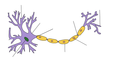
Schwann cell
This article needs additional citations for verification. (November 2010) |
| Schwann cell | |
|---|---|
 The PNS has satellite cells and Schwann cells. | |
| Identifiers | |
| MeSH | D012583 |
| FMA | 62121 |
| Anatomical terms of neuroanatomy | |
Schwann cells (named after physiologist Theodor Schwann) or neurolemmocytes are the principal glia of the peripheral nervous system (PNS). Glial cells function to support neurons and, in the PNS, also include satellite cells, olfactory ensheathing cells, enteric glia and glia that reside at sensory nerve endings, such as the Pacinian corpuscle. There are two types of Schwann cell, myelinating and nonmyelinating. Myelinating Schwann cells wrap around axons of motor and sensory neurons to form the myelin sheath.
Schwann cells are involved in many important aspects of peripheral nerve biology—the conduction of nervous impulses along axons, nerve development and regeneration, trophic support for neurons, production of the nerve extracellular matrix, modulation of neuromuscular synaptic activity, and presentation of antigens to T-lymphocytes.
Charcot–Marie–Tooth disease (CMT), Guillain–Barré syndrome (GBS, acute inflammatory demyelinating polyradiculopathy type), schwannomatosis, and chronic inflammatory demyelinating polyneuropathy (CIDP), and leprosy are all neuropathies involving Schwann cells.
Description

Schwann cells are a variety of glial cell that keep peripheral nerve fibres (both myelinated and unmyelinated) alive. In myelinated axons, Schwann cells form the myelin sheath (see above). The sheath is not continuous. Individual myelinating Schwann cells cover about 100 micrometres of an axon—equating to approximately 10,000 Schwann cells along a 1 metre length of the axon—which can be up to a metre or more in length. The gaps between adjacent Schwann cells are called nodes of Ranvier (see above). The vertebrate nervous system relies on the myelin sheath for insulation and as a method of decreasing membrane capacitance in the axon. The action potential jumps from node to node, in a process called saatory conduction, which can increase conduction velocity up to ten times, without an increase in axonal diameter. In this sense, Schwann cells are the peripheral nervous system's analogues of the central nervous system's oligodendrocytes. However, unlike oligodendrocytes, each myelinating Schwann cell provides insulation to only one axon (see image). This arrangement permits saltatory —conduction of action potentials with repropagation at the nodes of Ranvier. In this way, myelination greatly increases speed of conduction and saves energy.[1]
Non-myelinating Schwann cells are involved in maintenance of axons and are crucial for neuronal survival. Some group around smaller axons (External image here) and form Remak bundles. Myelinating Schwann cells begin to form the myelin sheath in mammals during fetal development and work by spiraling around the axon, sometimes with as many as 100 revolutions. A well-developed Schwann cell is shaped like a rolled-up sheet of paper, with layers of myelin in between each coil. The inner layers of the wrapping, which are predominantly membrane material, form the myelin sheath while the outermost layer of nucleated cytoplasm forms the neurolemma. Only a small volume of residual cytoplasm communicates the inner from the outer layers. This is seen histologically as the Schmidt-Lantermann incisure.
Schwann cell transplantation and regeneration
A number of experimental studies since 2001 have implanted Schwann cells in an attempt to induce remyelination in multiple sclerosis-afflicted patients.[2] In the past two decades, many studies have demonstrated positive results and potential for Schwann cell transplantation as a therapy for spinal cord injury, both in aiding regrowth and myelination of damaged CNS axons.[3] Schwann cell transplants in combination with other therapies such as Chondroitinase ABC have also been shown to be effective in functional recovery from spinal cord injury.[4] Indeed, Schwann cells are known for their roles in supporting nerve regeneration.[5] Nerves in the PNS consist of many axons myelinated by Schwann cells. If damage occurs to a nerve, the Schwann cells will aid in digestion of its axons (phagocytosis). Following this process, the Schwann cells can guide regeneration by forming a type of tunnel that leads toward the target neurons. The stump of the damaged axon is able to sprout, and those sprouts that grow through the Schwann-cell “tunnel” do so at the rate of approximately 1mm/day in good conditions. The rate of regeneration decreases with time. Successful axons can therefore reconnect with the muscles or organs they previously controlled with the help of Schwann cells, however, specificity is not maintained and errors are frequent, especially when long distances are involved.[6] Because of their ability to impact regeneration of axons, Schwann cells have been connected to preferential motor reinnervation as well. If Schwann cells are prevented from associating with axons, the axons die. Regenerating axons will not reach any target unless Schwann cells are there to support them and guide them. They have been shown to be in advance of the growth cones. Schwann cells are essential for the maintenance of healthy axons. They produce a variety of factors, including neurotrophins, and also transfer essential molecules across to axons.
Ganglioside
9-O-acetyl GD3 ganglioside is an acetylated glycolipid which is found in the cell membranes of many types of vertebrate cells. During peripheral nerve regeneration, 9-O-acetyl GD3 is expressed by Schwann cells.[7]
See also
References
- ^ Kalat, James W. Biological Psychology, 9th ed. USA: Thompson Learning, 2007.[page needed]
- ^ "First surgical transplant attempted to repair myelin". Inside MS. 2001.
- ^ Oudega, Martin; Xu, Xiao-Ming (2006). "Schwann Cell Transplantation for Repair of the Adult Spinal Cord". Journal of Neurotrauma. 23 (3–4): 453–67. doi:10.1089/neu.2006.23.453. PMID 16629629.
- ^ Fouad, Karim; Lisa Schnell; Mary B. Bunge; Martin E. Schwab; Thomas Liebscher; Damien D. Pearse (2 February 2005). "Combining Schwann Cell Bridges and Olfactory-Ensheathing Glia Grafts with Chondroitinase Promotes Locomotor Recovery after Complete Transection of the Spinal Cord". The Journal of Neuroscience. 25 (5): 1169–78. doi:10.1523/JNEUROSCI.3562-04.2005. PMID 15689553. Retrieved 15 May 2013.
- ^ Bhatheja, Kanav; Field, Jeffrey (2006). "Schwann cells: Origins and role in axonal maintenance and regeneration". The International Journal of Biochemistry & Cell Biology. 38 (12): 1995–9. doi:10.1016/j.biocel.2006.05.007. PMID 16807057.
- ^ Carlson, Neil R. Physiology of Behavior, 9th ed. USA: Pearson Education, Inc., 2007.[page needed]
- ^ Túlio Ribeiro-Resende, Victor; Lopes, Michelle (2010). "Involvement of 9-O-Acetyl GD3 Ganglioside in Mycobacterium leprae Infection of Schwann Cells" (PDF). J. Biol. Chem. 258. ASBMB: 34086–34096. doi:10.1074/jbc.M110.147272. Retrieved 14 April 2014.
{{cite journal}}: CS1 maint: unflagged free DOI (link)
External links
- Diagram at clc.uc.edu
- Histology image: 21301loa – Histology Learning System at Boston University—"Ultrastructure of the Cell: myelinated axon and Schwann cell"
- Cell Centered Database – Schwann cell

