Talk:Color vision
| This is the talk page for discussing improvements to the Color vision article. This is not a forum for general discussion of the article's subject. |
Article policies
|
| Find sources: Google (books · news · scholar · free images · WP refs) · FENS · JSTOR · TWL |
| Archives: 1Auto-archiving period: 2 months |
| This article has not yet been rated on Wikipedia's content assessment scale. It is of interest to the following WikiProjects: | |||||||||||||||||||||||||||||||||||||||||||||||||||||
Please add the quality rating to the {{WikiProject banner shell}} template instead of this project banner. See WP:PIQA for details.
Please add the quality rating to the {{WikiProject banner shell}} template instead of this project banner. See WP:PIQA for details.
Please add the quality rating to the {{WikiProject banner shell}} template instead of this project banner. See WP:PIQA for details.
Please add the quality rating to the {{WikiProject banner shell}} template instead of this project banner. See WP:PIQA for details.
| |||||||||||||||||||||||||||||||||||||||||||||||||||||
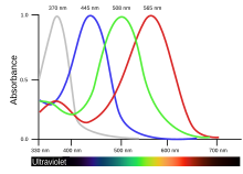
The second (violet) maximum of the sensitivity of the red cones
(I recently started this topic, but after that all the talks from 2006 to 2019 ended by my message were automatically archived.)

In some sites (e.g. https://midimagic.sgc-hosting.com/huvision.htm) I see charts of light sensitivity curves looking different. Namely, the curve of the red-sensitive cones has a second (tiny) peak in the violet range. It is said:
The erythropsin in the red-sensitive cones is sensitive to two ranges of wavelengths. The major range is between 500 nm and 760 nm, peaking at 600 nm. This includes green, yellow, orange, and red light. The minor range is between 380 nm and 450 nm, peaking at 420 nm. This includes violet and some blue. The minor range is what makes the hues appear to form a circle instead of a straight line.
There is no reference to scientific papers neither numerical tables in https://midimagic.sgc-hosting.com/huvision.htm. I have not found any evidence of that in Wikipedia except that of a bird's cone cells rather than human's (see the chart):
Is this a fake science or an obsolete disproved theory? If not, could anybody find a reference to an authoritative source?Ufim (talk) 08:37, 28 April 2019 (UTC)
- ^ Figure data, uncorrected absorbance curve fits, from Hart, NS; Partridge, JC; Bennett, ATD; Cuthill, IC (2000). "Visual pigments, cone oil droplets and ocular media in four species of estrildid finch". Journal of Comparative Physiology A. 186 (7–8): 681–694. doi:10.1007/s003590000121.
- B-Class color articles
- Top-importance color articles
- All WikiProject Color pages
- C-Class Biology articles
- High-importance Biology articles
- WikiProject Biology articles
- C-Class neuroscience articles
- High-importance neuroscience articles
- B-Class psychology articles
- High-importance psychology articles
- WikiProject Psychology articles

