Coronary arteries
| Coronary arteries | |
|---|---|
 Coronary arteries (labeled in red text) and other major landmarks (in blue text) | |
| Identifiers | |
| FMA | 49893 |
| Anatomical terminology | |
The coronary arteries are the blood vessels (arteries) of coronary circulation, which transports oxygenated blood to the actual heart muscle. The heart requires a continuous supply of oxygen to function and survive, much like any other tissue or organ of the body. [1]
The coronary arteries wrap around the entire heart. The two main branches are the left coronary artery (LCA) and right coronary artery (RCA). The arteries can additionally be categorized based on the area of the heart they innervate. These categories are called epicardial (above the epicardium or the outer surface of the heart) and microvascular (close to the endocardium or the innermost tissue of the heart).[2]
Reduced function of the coronary arteries can lead to decreased flow of oxygen and nutrients to the heart. Not only does this affect supply to the heart muscle itself, but it also can affect the ability of the heart to pump blood throughout the body. Therefore, any disorder or disease to our coronary arteries can have a serious impacts on our health, possibly leading to angina, a heart attack, and even death. [3]
Structure
The coronary arteries are mainly composed of the left and right coronary arteries, both of which give off several branches as shown in the 'Coronary artery flow' figure.

- Left coronary artery (LCA)
- Right coronary artery (RCA)
The left coronary artery (LCA) arises from the aorta within the left cusp of the aortic valve and feeds blood to the left side of the heart. It branches into two arteries, the left anterior descending and the left circumflex. The left anterior descending artery perfuses the interventricular septum and anterior wall of the left ventricle. The left circumflex artery perfuses the left ventricular free wall. In approximately 33% of individuals, the left coronary artery gives rise to the posterior descending artery[4] which perfuses the posterior and inferior walls of the left ventricle. Sometimes a third branch is formed at the fork between left anterior descending and left circumflex arteries known as a ramus or intermediate artery.[5]
The right coronary artery (RCA) originates within the right cusp of the aortic valve. It travels down the right coronary sulcus, towards the crux of the heart. The RCA primarily branches into the right marginal arteries, and in 67% of individuals gives rise to the posterior descending artery[4]. The right marginal arteries perfuse the right ventricle and the posterior descending artery perfuses the left ventricular posterior and inferior walls.
There is also the conus artery, which is only present in about 45 per cent of the human population, and which may provide collateral blood flow to the heart when the left anterior descending artery is occluded.[6][7]
Clinical significance
Either or both arteriosclerosis and atherosclerosis, can cause one or more of the coronary arteries or their branches to become seriously blocked, leading to angina, heart attack, or both.[8] Percutaneous coronary interventions (such as balloon angioplasty) or coronary artery bypass surgery can be performed to decrease or bypass the blockages (respectively).[9]
The coronary arteries can constrict as a response to various stimuli, mostly chemical. This is known as a coronary reflex.
There is also a rare condition known as spontaneous coronary artery dissection.
Name etymology
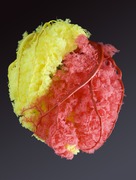
The word corona is a Latin word meaning "crown", from the Ancient Greek κορώνη (korōnè, “garland, wreath”). It was applied to the coronary arteries because of a notional resemblance (compare the photos).
Additional images
References
- ^ "Coronary Arteries". Texas Heart Institute. Retrieved 2019-09-01.
- ^ Petersen, J. W.; Pepine, C. J. (2014). "Microvascular Coronary Dysfunction and Ischemic Heart Disease – Where Are We in 2014?". Trends in Cardiovascular Medicine. 25 (2): 98–103. doi:10.1016/j.tcm.2014.09.013. PMC 4336803. PMID 25454903.
- ^ "Anatomy and Function of the Coronary Arteries". www.hopkinsmedicine.org. Retrieved 2019-09-01.
- ^ a b Costanzo, Linda S. (2018). Physiology (6th ed.). Philadelphia, PA: Elsevier. ISBN 9780323511896. OCLC 965761862.
- ^ Fuster, V; Alexander RW; O'Rourke RA (2001). Hurst's The Heart (10th ed.). McGraw-Hill. p. 53. ISBN 978-0-07-135694-7.
- ^ Wynn GJ, Noronha B, Burgess MI (2008). "Functional significance of the conus artery as a collateral to an occluded left anterior descending artery demonstrated by stress echocardiography". International Journal of Cardiology. 140 (1): e14–5. doi:10.1016/j.ijcard.2008.11.039. PMID 19108914.
- ^ Schlesinger MJ, Zoll PM, Wessler S (1949). "The conus artery: a third coronary artery". American Heart Journal. 38 (6): 823–38. doi:10.1016/0002-8703(49)90884-4. PMID 15395916.
- ^ Kumar, Vinay; Abbas, Abul K.; Aster, Jon C., eds. (2015). Robbins and Cotran Pathologic Basis of Disease. Illustrated by Perkins, James A. (9th ed.). Philadelphia, PA: Saunders. ISBN 9781455726134. OCLC 879416939.
- ^ Lilly, Leonard S. (2011). Pathophysiology of heart disease: a collaborative project of medical students and faculty (5th ed.). Baltimore, MD: Wolters Kluwer/Lippincott Williams & Wilkins. ISBN 9781605477237. OCLC 649701807.


