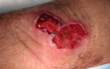
Mycobacterium ulcerans
| Mycobacterium ulcerans | |
|---|---|

| |
| Scientific classification | |
| Kingdom: | |
| Phylum: | |
| Order: | |
| Suborder: | |
| Family: | |
| Genus: | |
| Species: | M. ulcerans
|
| Binomial name | |
| Mycobacterium ulcerans MacCallum et al. 1950
| |
Description
Under a microscope, M. ulcerans appear as rods.[1] They appear purple ("Gram positive") under Gram stain and bright red ("acid fast") under Ziehl–Neelsen stain.[1] On laboratory media, M. ulcerans grow slowly, forming small transparent colonies after four weeks.[1] As colonies age, they develop irregular outlines and a rough, yellow surface.[1]
Evolution
M. ulcerans likely evolved from the closely related aquatic pathogen Mycobacterium marinum around one million years ago.[2] The two species are genetically very similar, and have identical 16S ribosomal RNA genes.[1] However relative to M. marinum, M. ulcerans has undergone substantial genome reduction, shedding over a thousand kilobases of genetic content including nearly 1300 genes (23% of the total M. marinum genes) and sustaining the inactivation of an additional 700 genes.[3] Some of these genes were inactivated by the proliferation of two mobile genetic elements, called "IS2404" (213 copies) and "IS2606" (91 copies), neither of which are present in M. marinum.[3] Additionally, M. ulcerans has acquired a 174 kilobase plasmid, termed "pMUM001", which is involved in the production of the toxin mycolactone.[3] Other closely related mycobacteria produce mycolactone and infect various aquatic animals; these are sometimes described as distinct species (M. pseudoshottsii, M. liflandii, M. shinshuense and sometimes M. marinum) and sometimes as different lineages of M. ulcerans. Regardless, all mycolactone-producing mycobacteria share a common ancestor distinct from non-mycolactone-producing M. marinum.[4]
References
- ^ a b c d e Magee & Ward 2015, pp. 28–29.
- ^ Röltgen & Pluschke 2019, pp. 4–5.
- ^ a b c Demangel, Stinear & Cole 2009, pp. 52, 54.
- ^ Vandelannoote et al. 2019, pp. 108–109.
Works cited
- Demangel C, Stinear TP, Cole ST (2009). "Buruli ulcer: reductive evolution enhances pathogeneicity of Mycobacterium ulcerans". Nature Reviews Microbiology. 7: 50–60. doi:10.1038/nrmicro2077.
- Magee JG, Ward AC (September 2015). "Mycobacterium". In Whitman WB (ed.). Bergey's Manual of Systematics of Archaea and Bacteria. John Wiley & Sons, Bergey's Manual Trust. doi:10.1002/9781118960608.gbm00029. ISBN 9781118960608.
- Röltgen K, Pluschke G (2019). "Buruli Ulcer: History and Disease Burden". In Röltgen K, Pluschke G (eds.). Buruli Ulcer: Mycobacterium Ulcerans Disease. Cham: Springer. PMID 32091710.
- Vandelannoote K, Eddyani M, Buultjens A, Stinear TP (2019). "Population Genomics and Molecular Epidemiology of Mycobacterium ulcerans". In Röltgen K, Pluschke G (eds.). Buruli Ulcer: Mycobacterium Ulcerans Disease. Cham: Springer. PMID 32091703.
External links
- "Mycobacterium ulcerans". NCBI Taxonomy Browser. 1809.
