谷胱甘肽还原酶
外观
此條目需要擴充。 (2011年9月15日) |
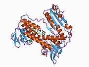
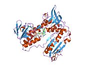
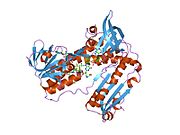
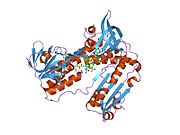
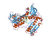
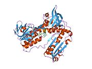
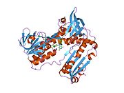
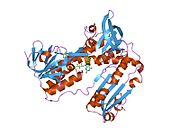
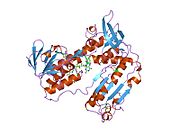
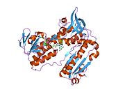
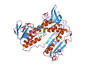
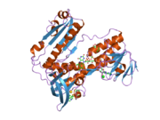
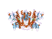
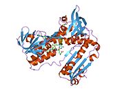
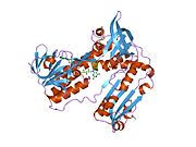
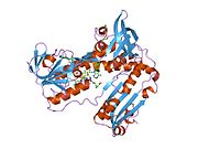
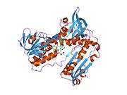
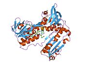
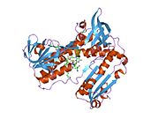
谷胱甘肽还原酶[1](英語:Glutathione reductase,EC 1.8.1.7)是一种将氧化型谷胱甘肽(GSSG)还原成为硫醇型谷胱甘肽的酶,而谷胱甘肽是一种重要的细胞抗氧化剂[2][3]。
对于一摩尔的氧化型谷胱甘肽(GSSG),需要一摩尔的NADPH将氧化型谷胱甘肽还原为还原型谷胱甘肽。这种酶形成一个FAD结合的同型二聚体。谷胱甘肽还原酶在所有界中都是进化保守的。在细菌、酵母与动物中,编码谷胱甘肽还原酶的基因是同一种;然而,在植物基因组中,有两个被编码的谷胱甘肽还原酶。果蝇与锥体虫甚至都没有编码谷胱甘肽还原酶的基因[4]。在这些生物中,谷胱甘肽还原酶的还原是分别由硫氧还蛋白以及锥虫胱甘肽系统来完成的[4][5]。
以NADPH为氢供体、氧化型谷胱甘肽为氢受体的还原酶,反应生成NADP+和还原型谷胱甘肽。
参考文献
[编辑]- ^ [1][永久失效連結]
- ^ Meister A. Glutathione metabolism and its selective modification. J. Biol. Chem. 1988, 263 (33): 17205–8 [2011-09-19]. PMID 3053703. (原始内容存档于2009-08-17).
- ^ Mannervik B. The enzymes of glutathione metabolism: an overview. Biochem. Soc. Trans. 1987, 15 (4): 717–8. PMID 3315772.
- ^ 4.0 4.1 Kanzok SM, Fechner A, Bauer H, Ulschmid JK, Müller HM, Botella-Munoz J, Schneuwly S, Schirmer R, Becker K. Substitution of the thioredoxin system for glutathione reductase in Drosophila melanogaster. Science. 2001, 291 (5504): 643–6. PMID 11158675. doi:10.1126/science.291.5504.643.
- ^ Krauth-Siegel RL, Comini MA. Redox control in trypanosomatids, parasitic protozoa with trypanothione-based thiol metabolism. Biochim Biophys Acta. 2008, 1780 (11): 1236–48. PMID 18395526. doi:10.1016/j.bbagen.2008.03.006.
深入阅读
[编辑]- Sinet PM, Bresson JL, Couturier J; et al. [Possible localization of the glutathione reductase (EC 1.6.4.2) on the 8p21 band]. Ann. Genet. 1977, 20 (1): 13–7. PMID 302667.
- Krohne-Ehrich G, Schirmer RH, Untucht-Grau R. Glutathione reductase from human erythrocytes. Isolation of the enzyme and sequence analysis of the redox-active peptide.. Eur. J. Biochem. 1978, 80 (1): 65–71. PMID 923580. doi:10.1111/j.1432-1033.1977.tb11856.x.
- Loos H, Roos D, Weening R, Houwerzijl J. Familial deficiency of glutathione reductase in human blood cells.. Blood. 1976, 48 (1): 53–62. PMID 947404.
- Tutic M, Lu XA, Schirmer RH, Werner D. Cloning and sequencing of mammalian glutathione reductase cDNA.. Eur. J. Biochem. 1990, 188 (3): 523–8. PMID 2185014. doi:10.1111/j.1432-1033.1990.tb15431.x.
- Palmer EJ, MacManus JP, Mutus B. Inhibition of glutathione reductase by oncomodulin.. Arch. Biochem. Biophys. 1990, 277 (1): 149–54. PMID 2306116. doi:10.1016/0003-9861(90)90563-E.
- Arnold HH, Heinze H. Treatment of human peripheral lymphocytes with concanavalin A activates expression of glutathione reductase.. FEBS Lett. 1990, 267 (2): 189–92. PMID 2379581. doi:10.1016/0014-5793(90)80922-6.
- Karplus PA, Schulz GE. Refined structure of glutathione reductase at 1.54 A resolution.. J. Mol. Biol. 1987, 195 (3): 701–29. PMID 3656429. doi:10.1016/0022-2836(87)90191-4.
- Pai EF, Schulz GE. The catalytic mechanism of glutathione reductase as derived from x-ray diffraction analyses of reaction intermediates.. J. Biol. Chem. 1983, 258 (3): 1752–7. PMID 6822532.
- Krauth-Siegel RL, Blatterspiel R, Saleh M; et al. Glutathione reductase from human erythrocytes. The sequences of the NADPH domain and of the interface domain.. Eur. J. Biochem. 1982, 121 (2): 259–67. PMID 7060551. doi:10.1111/j.1432-1033.1982.tb05780.x.
- Thieme R, Pai EF, Schirmer RH, Schulz GE. Three-dimensional structure of glutathione reductase at 2 A resolution.. J. Mol. Biol. 1982, 152 (4): 763–82. PMID 7334521. doi:10.1016/0022-2836(81)90126-1.
- Huang J, Philbert MA. Distribution of glutathione and glutathione-related enzyme systems in mitochondria and cytosol of cultured cerebellar astrocytes and granule cells.. Brain Res. 1995, 680 (1–2): 16–22. PMID 7663973. doi:10.1016/0006-8993(95)00209-9.
- Savvides SN, Karplus PA. Kinetics and crystallographic analysis of human glutathione reductase in complex with a xanthene inhibitor. J. Biol. Chem. 1996, 271 (14): 8101–7. PMID 8626496. doi:10.1074/jbc.271.14.8101.
- Nordhoff A, Tziatzios C, van den Broek JA; et al. Denaturation and reactivation of dimeric human glutathione reductase--an assay for folding inhibitors. Eur. J. Biochem. 1997, 245 (2): 273–82. PMID 9151953. doi:10.1111/j.1432-1033.1997.00273.x.
- Stoll VS, Simpson SJ, Krauth-Siegel RL; et al. Glutathione reductase turned into trypanothione reductase: structural analysis of an engineered change in substrate specificity. Biochemistry. 1997, 36 (21): 6437–47. PMID 9174360. doi:10.1021/bi963074p.
- Becker K, Savvides SN, Keese M; et al. Enzyme inactivation through sulfhydryl oxidation by physiologic NO-carriers. Nat. Struct. Biol. 1998, 5 (4): 267–71. PMID 9546215. doi:10.1038/nsb0498-267.
- Kelner MJ, Montoya MA. Structural organization of the human glutathione reductase gene: determination of correct cDNA sequence and identification of a mitochondrial leader sequence. Biochem. Biophys. Res. Commun. 2000, 269 (2): 366–8. PMID 10708558. doi:10.1006/bbrc.2000.2267.
- Qanungo S, Mukherjea M. Ontogenic profile of some antioxidants and lipid peroxidation in human placental and fetal tissues. Mol. Cell. Biochem. 2001, 215 (1–2): 11–9. PMID 11204445. doi:10.1023/A:1026511420505.
- Berry Y, Truscott RJ. The presence of a human UV filter within the lens represents an oxidative stress. Exp. Eye Res. 2001, 72 (4): 411–21. PMID 11273669. doi:10.1006/exer.2000.0970.
- Rhie G, Shin MH, Seo JY; et al. Aging- and photoaging-dependent changes of enzymic and nonenzymic antioxidants in the epidermis and dermis of human skin in vivo. J. Invest. Dermatol. 2001, 117 (5): 1212–7. PMID 11710935. doi:10.1046/j.0022-202x.2001.01469.x.
- Zatorska A, Józwiak Z. Involvement of glutathione and glutathione-related enzymes in the protection of normal and trisomic human fibroblasts against daunorubicin. Cell Biol. Int. 2003, 26 (5): 383–91. PMID 12095224. doi:10.1006/cbir.2002.0861.


